. 2023 Dec 23:16:3699-3711.
doi: 10.2147/CCID.S439760. eCollection 2023.

Adipose-Derived Stem Cell Exosomes Alleviate Psoriasis Serum Exosomes-Induced Inflammation by Regulating Autophagy and Redox Status in Keratinocytes

Affiliations

Adipose-Derived Stem Cell Exosomes Alleviate Psoriasis Serum Exosomes-Induced Inflammation by Regulating Autophagy and Redox Status in Keratinocytes

Hye Ran Kim et al. Clin Cosmet Investig Dermatol. .

Abstract

Introduction: Exosomes play a key role in cell communication and are involved in both pathological and physiological processes. Autophagy dysfunction and oxidative stress are linked to immune-mediated inflammatory diseases such as psoriasis. Stem cell-derived exosomes exhibit immunomodulatory and antioxidant efficacy.

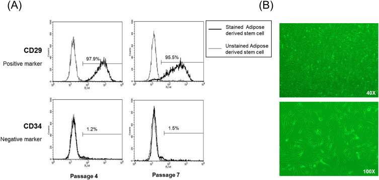
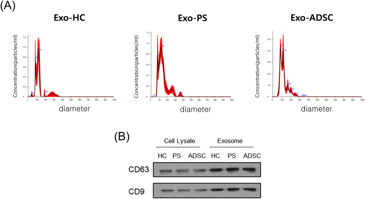
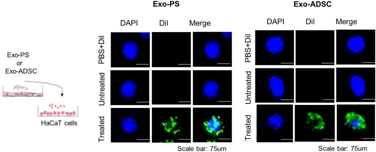
Methods: We aimed to investigate the impact of psoriasis serum-derived exosomes on inflammation, oxidative stress, and autophagy in keratinocytes. Additionally, we explored the therapeutic potential of adipose-derived stem cell (ADSC) exosomes against inflammation induced by psoriasis serum exosomes. To validate psoriasis patient serum-derived exosomes and ADSC exosomes, we used nanoparticle tracking analysis, Western blotting, flow cytometry, and immunofluorescence. qPCR was used to study changes in the gene expression of proinflammatory cytokines and oxidative stress markers in HaCaT cells treated with psoriasis serum-derived exosomes or ADSC exosomes. The effects of these exosomes on autophagy in HaCaT cells were evaluated by Western blotting and immunofluorescence.

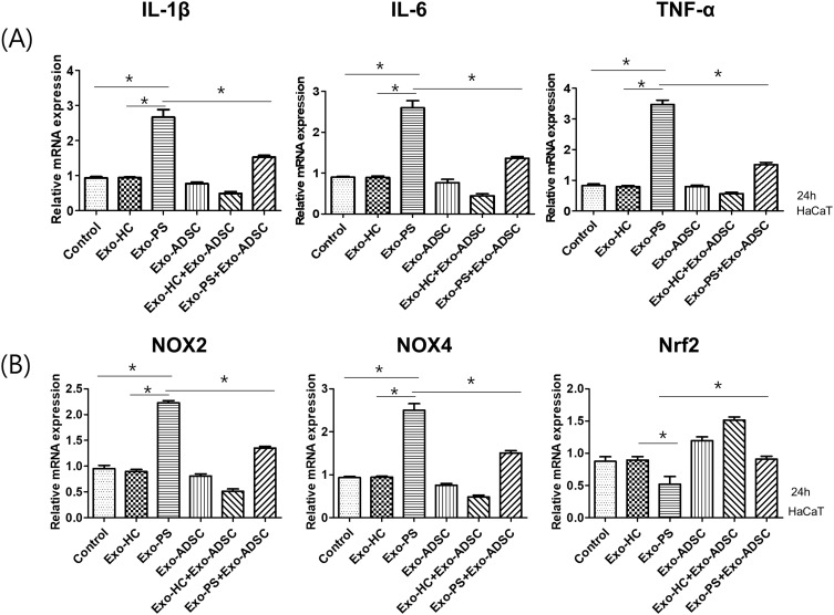
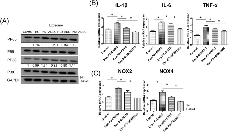
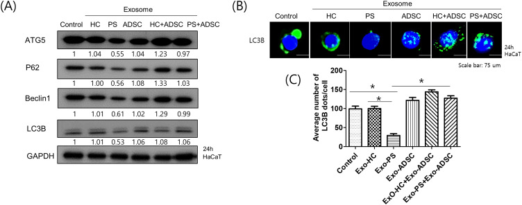
Result: The treatment of HaCaT cells with psoriasis serum-derived exosomes increased proinflammatory cytokine production and oxidative stress-related factor (Nox2 and Nox4) expression and decreased Nrf2 expression via P65/NF-κB and P38/MAPK activation. Compared with healthy control serum-derived exosomes, psoriasis serum-derived exosomes decreased ATG5, P62, Beclin1, and LC3 expression and autophagosome production in HaCaT cells. Conversely, ADSC exosomes suppressed proinflammatory cytokine and oxidative stress production, and restored autophagy in HaCaT cells treated with psoriasis serum-derived exosomes.

Discussion: These findings suggest that ADSC exosomes exhibit a suppressive effect on psoriasis serum exosome-induced inflammation and oxidative stress by regulating autophagy in keratinocytes.

Keywords: adipose-derived stem cell; autophagy; exosomes; oxidative stress; psoriasis.


Conflict of interest statement

The authors report no conflicts of interest in this work.

Figures

Figure 1
Figure 1
Characterization of ADSCs. (A) Assessment of cell surface markers by flow cytometry. ADSCs were positive for the MSC marker CD29 but negative for the endothelial marker CD34. (B) Fibroblast-like morphology of ADSCs (40X, 100X).
Figure 2
Figure 2
Characterization of exosomes (A) Results of nanoparticle tracking analysis demonstrating a similar size distribution in separated exosomes from healthy control serum or psoriasis patient serum or ADSCs (n = 4). (B) The representative exosome markers CD63 and CD9 were detected by Western blotting (n = 3).
Figure 3
Figure 3
Schematic of the protocol for exosome uptake analysis and representative immunofluorescence images of HaCaT cells treated with psoriasis patient serum or ADSC-derived exosomes. Exosomes were stained with the fluorescent green dye DiI, and HaCaT cells were incubated with psoriasis serum or ADSC-derived exosomes for 2 h. Nuclei were counterstained with blue. Scale bar, 75 µm.
Figure 4
Figure 4
The effects of exosomes on the production of proinflammatory cytokines and oxidative stress-related factors. (A) The expression of IL-1β, IL-6, and TNF-α mRNA in exosome-treated HaCaT cells. (B) The expression of NOX2, NOX4, and Nrf2 mRNA in exosome-treated HaCaT cells. Statistical significance was determined by the one-way analysis of variance with Tukey’s post-hoc test. Data represent the mean ± S.D. of 3 independent experiments. *P < 0.05.
Figure 5
Figure 5
Psoriasis serum-derived exosomes induced proinflammatory cytokine and oxidative stress production via P65/NF-κB and P38/MAPK signaling. (A) The phosphorylation levels of P65 and P38 in exosome-treated HaCaT cells detected by Western blot. The relative expression was normalized to GAPDH. The results are representative of three independent experiments. HC, healthy control serum; PS, psoriasis patient serum; ADSCs, adipose-derived stem cells; GAPDH, glyceraldehyde 3-phosphate dehydrogenase. (B and C) The mRNA expression of IL-1β, IL-6, TNF-α, NOX2, and NOX4 in HaCaT cells pretreated with or without P65 or P38 inhibitors and treated with psoriasis serum-derived exosomes. HaCaT cells were pretreated with chemical inhibitors for P65 [pyrrolidine dithiocarbamate (PDTC)] and P38 (SB203580) for 1 h and then stimulated by PS exosomes for 24 h, followed by qPCR for genes including IL-1β, IL-6, TNF-α, NOX2, and NOX4. Statistical significance was determined by the one-way analysis of variance with Tukey’s post-hoc test. Data represent the mean ± S.D. of 3 independent experiments. *P < 0.05.
Figure 6
Figure 6
The effects of exosomes on autophagy in HaCaT cells. (A) The effects of exosomes on autophagy-related factor expression in HaCaT cells. HaCaT cells were stimulated with exosomes derived from HC, PS, and ADSCs for 24 h and then analyzed by Western blotting using antibodies against ATG5, P62, Beclin1 and LC3B; relative expression was normalized to GAPDH. The results are representative of three independent experiments. (B) Representative images of immunofluorescence staining of LC3B. Immunofluorescence staining to assess the autophagy marker LC3B in exosome-treated HaCaT cells. Scale bars, 75 μm. (C) The relative quantification of LC3B based on the average fluorescence intensity per cell is shown. Each comprising at least 250 cells scored in five random fields. Statistical significance was determined by the one-way analysis of variance with Tukey’s post-hoc test. Data represent the mean ± S.D. of 3 independent experiments. *P < 0.05.

Similar articles

Cited by

References

    1. Chhabra S, Dogra S, Sharma K, Raychaudhuri SK, Raychaudhuri SP. Recent update on immunopathogenesis of psoriasis. Indian J Dermatol. 2022;67(4):360–373. doi:10.4103/ijd.ijd_569_22 - DOI - PMC - PubMed
    1. Takeshita J, Grewal S, Langan SM, et al. Psoriasis and comorbid diseases: epidemiology. J Am Acad Dermatol. 2017;76(3):377–390. doi:10.1016/j.jaad.2016.07.064 - DOI - PMC - PubMed
    1. Gelfand JM, Feldman SR, Stern RS, Thomas J, Rolstad T, Margolis DJ. Determinants of quality of life in patients with psoriasis: a study from the US population. J Am Acad Dermatol. 2004;51(5):704–708. doi:10.1016/j.jaad.2004.04.014 - DOI - PubMed
    1. Plenkowska J, Gabig-Ciminska M, Mozolewski P. Oxidative stress as an important contributor to the pathogenesis of psoriasis. Int J Mol Sci. 2020;21(17):6206. - PMC - PubMed
    1. Arthur JS, Darragh J. Signaling downstream of p38 in psoriasis. J Invest Dermatol. 2006;126(8):1689–1691. doi:10.1038/sj.jid.5700280 - DOI - PubMed