

Case Reports
doi: 10.1016/j.xjtc.2023.09.026.
eCollection 2023 Dec.
Interventricular septal dissection with perforations following takotsubo cardiomyopathy
Affiliations
- PMID: 38152206
- PMCID: PMC10750958
- DOI: 10.1016/j.xjtc.2023.09.026
Item in Clipboard
Case Reports
Interventricular septal dissection with perforations following takotsubo cardiomyopathy
JTCVS Tech.
.
No abstract available
Conflict of interest statement
The authors reported no conflicts of interest. The Journal policy requires editors and reviewers to disclose conflicts of interest and to decline handling or reviewing manuscripts for which they may have a conflict of interest. The editors and reviewers of this article have no conflicts of interest.
Figures
Ventricular septal perforation with dissection following takotsubo cardiomyopathy.
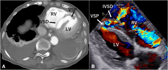
A, Contrast-enhanced CT demonstrating extensive IVSD complicated with VSP. B, Transthoracic echocardiography showing the dissection of the IVS in color Doppler indicating plural flows through the VSP, near the apex. VSP, Ventricular septal perforation; RV, right ventricle; IVSD, interventricular septal dissection; LV, left ventricle.
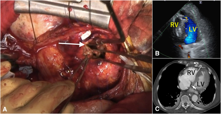
A, Intraoperative image of a single tear (1 cm × 1 cm) of the ventricular septal wall just under the incision (arrow). The left ventricular septum around the ostium of the dissection tract is not fragile and reveals no ischemic change. B, Postoperative echocardiography showing no shunt after VSP repair. C, Contrast-enhanced CT showing successful VSP repair. RV, Right ventricle; LV, left ventricle.
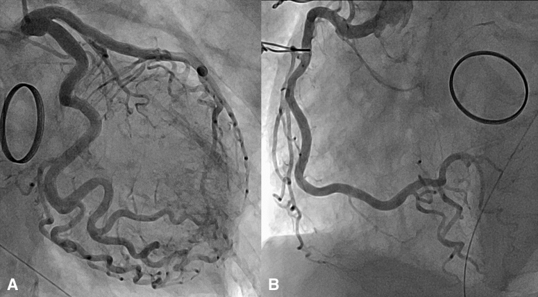
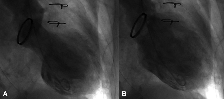
Coronary angiography of (A) the left coronary artery and (B) the right coronary artery revealing no lesions.
Left ventriculography of (A) systole and (B) diastole showing apical akinesis.
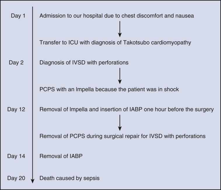
Sequence of events from the admission to postoperative death. ICU, Intensive care unit; IVSD, interventricular septal dissection; PCPS, percutaneous cardiopulmonary support; IABP, intra-aortic balloon pump.
References
-
- Mariscalco G., Cattaneo P., Rossi A., Baravelli M., Piffaretti G., Scannapieco A., et al. Tako-tsubo cardiomyopathy complicated by ventricular septal perforation and septal dissection. Heart Vessels. 2010;25:73–75. - PubMed
-
- Isoda S., Osako M., Kimura T., Mashiko Y., Yamanaka N., Nakamura S., et al. Midterm results of the “sandwich technique” via a right ventricle incision to repair post-infarction ventricular septal defect. Ann Thorac Cardiovasc Surg. 2012;18:318–321. - PubMed
-
- Miyake K., Funatsu T., Kondoh H., Taniguchi K. Rare complication of takotsubo cardiomyopathy: ventricular septal perforation with septal dissection. J Card Surg. 2016;31:150–153. - PubMed
-
- Furui M., Sakurai Y., Kakii B., Asanuma M., Nishioka H., Yoshida T. Benefits and risks of delayed surgery for ventricular septal rupture after acute myocardial infarction. Int Heart J. 2022;63:433–440. - PubMed
Publication types
LinkOut - more resources
Full Text Sources
