. 2023 Dec 12:14:1326160.
doi: 10.3389/fphys.2023.1326160. eCollection 2023.

Injection of IK1 through dynamic clamp can make all the difference in patch-clamp studies on hiPSC-derived cardiomyocytes

Affiliations

Injection of IK1 through dynamic clamp can make all the difference in patch-clamp studies on hiPSC-derived cardiomyocytes

Arie O Verkerk et al. Front Physiol. .

Abstract

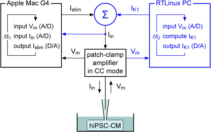
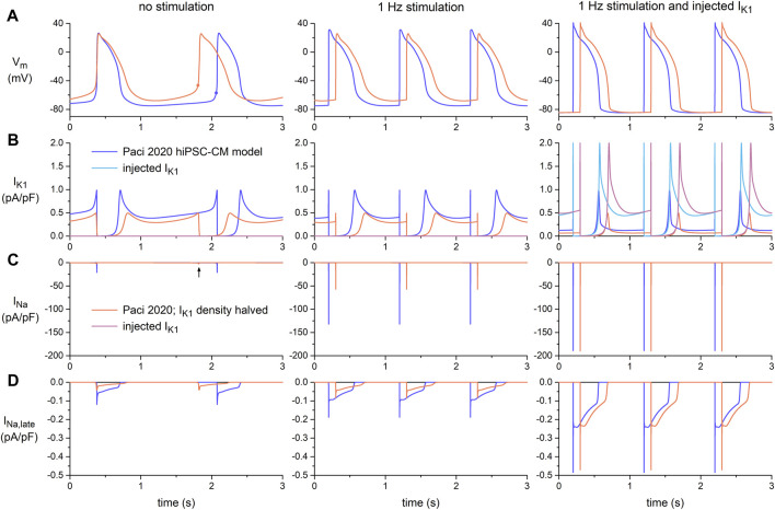
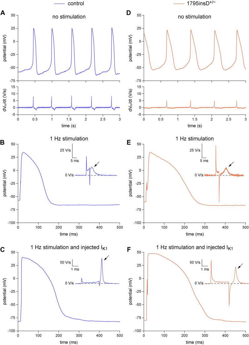
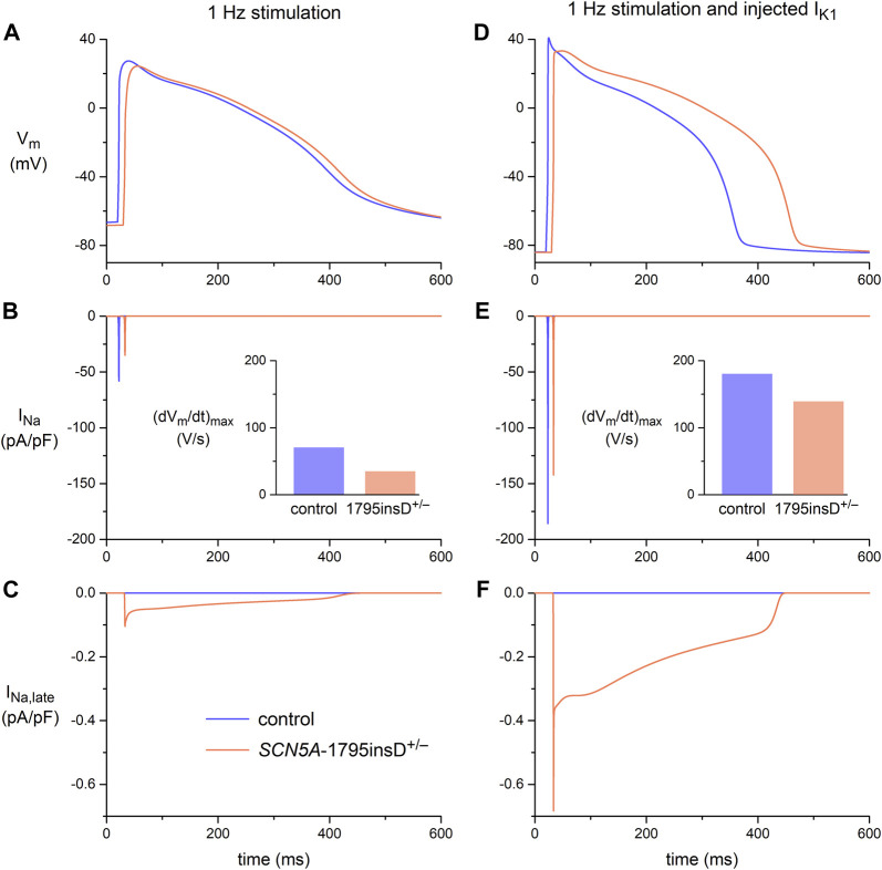
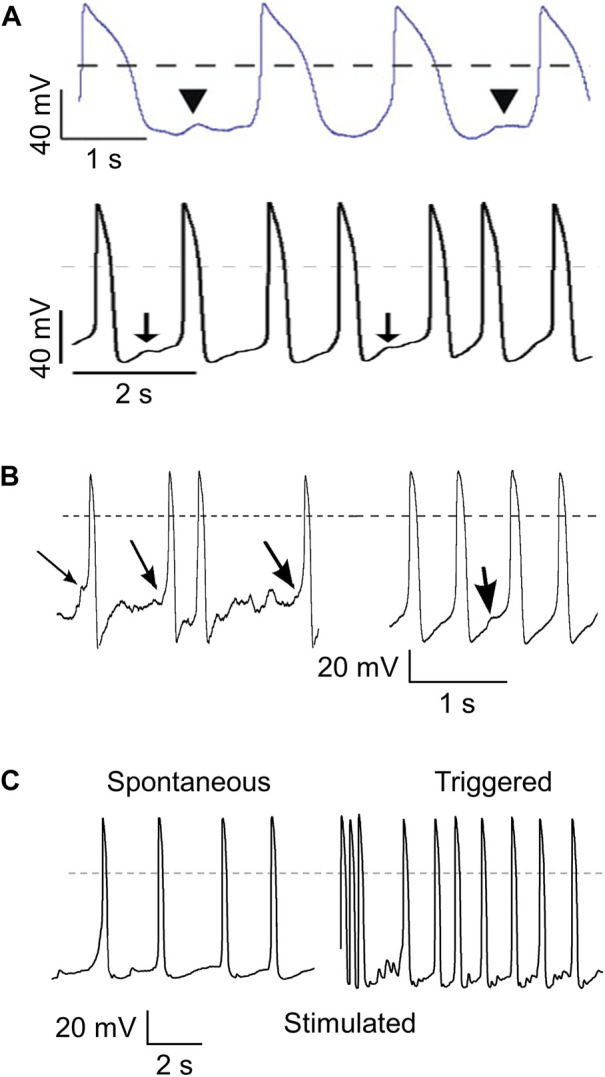
Human-induced stem cell-derived cardiomyocytes (hiPSC-CMs) are a valuable tool for studying development, pharmacology, and (inherited) arrhythmias. Unfortunately, hiPSC-CMs are depolarized and spontaneously active, even the working cardiomyocyte subtypes such as atrial- and ventricular-like hiPSC-CMs, in contrast to the situation in the atria and ventricles of adult human hearts. Great efforts have been made, using many different strategies, to generate more mature, quiescent hiPSC-CMs with more close-to-physiological resting membrane potentials, but despite promising results, it is still difficult to obtain hiPSC-CMs with such properties. The dynamic clamp technique allows to inject a current with characteristics of the inward rectifier potassium current (IK1), computed in real time according to the actual membrane potential, into patch-clamped hiPSC-CMs during action potential measurements. This results in quiescent hiPSC-CMs with a close-to-physiological resting membrane potential. As a result, action potential measurements can be performed with normal ion channel availability, which is particularly important for the physiological functioning of the cardiac SCN5A-encoded fast sodium current (INa). We performed in vitro and in silico experiments to assess the beneficial effects of the dynamic clamp technique in dissecting the functional consequences of the SCN5A-1795insD+/- mutation. In two separate sets of patch-clamp experiments on control hiPSC-CMs and on hiPSC-CMs with mutations in ACADVL and GNB5, we assessed the value of dynamic clamp in detecting delayed afterdepolarizations and in investigating factors that modulate the resting membrane potential. We conclude that the dynamic clamp technique has highly beneficial effects in all of the aforementioned settings and should be widely used in patch-clamp studies on hiPSC-CMs while waiting for the ultimate fully mature hiPSC-CMs.

Keywords: GNB5; SCN5A; acetylcholine-activated potassium current; delayed afterdepolarizations; fast sodium current; inward rectifier potassium current; triggered action potentials; ventricular action potential.


Conflict of interest statement

The authors declare that the research was conducted in the absence of any commercial or financial relationships that could be construed as a potential conflict of interest.

Figures

FIGURE 1
FIGURE 1
Dynamic clamp setup. The dynamic clamp component (represented by the blue section on the right) supplements the standard patch-clamp configuration (represented in black). The synthetic inward rectifier K+ current (IK1) is computed in real time by a Real-Time Linux (RTLinux) based PC in response to the recorded membrane potential (Vm) and then added to any stimulus current (Istim). The resulting composite current (Iin) is then transferred to the patch-clamp amplifier, which operates in current clamp (CC) mode and injects Iin into the patched hiPSC-CM. The IK1 and Istim signals can be combined in a separate electronic box (Σ) or within the patch clamp amplifier itself, based on the amplifier input options. This process is updated with a time step Δt2. Istim is sent out by the Apple Macintosh (Mac) G4 computer (or any other computer) that runs the regular patch-clamp software and is used to control the experiment. It records both Vm and Iin with a time step Δt1. Because Iin is read in, the latter computer contains all the data required for offline analysis, as in a regular patch clamp experiment.
FIGURE 2
FIGURE 2
Amplitude of the inward rectifier K+ current (IK1) in isolated human ventricular cardiomyocytes and in hiPSC-CMs, and current-voltage relationship of IK1 used in dynamic clamp experiments. (A) Peak outward amplitude of IK1 in studies on isolated human ventricular cardiomyocytes (top) and in studies on hiPSC-CMs (bottom). RT: room temperature; [K+]e: extracellular K+ concentration; ML: cultured as monolayer; EHT: cultured as 3D engineered heart tissue. (B) Current-voltage relationships of IK1 used in the dynamic clamp studies of Meijer van Putten et al. (2015) and Altomare et al. (2023), and in the Paci2020 model of a ventricular-like hiPSC-CM (solid traces). The dotted trace shows the steady-state current-voltage relationship of the hyperpolarization-activated ‘funny current’ (If) of the Paci2020 model.
FIGURE 3
FIGURE 3
Electrical activity in the Paci2020 model of a ventricular-like hiPSC-CM during spontaneous activity (no stimulation; left panels), during 1 Hz stimulation (middle panels), and during 1 Hz stimulation combined with the simulated injection of the IK1 of Meijer van Putten et al. (2015) (right panels, reproduced with permission). The current-voltage relationship of this injected IK1 is shown in Figure 2B. The blue traces are obtained with the original native IK1 of the Paci2020 model and the orange ones with the current density of this native IK1 halved. (A) Membrane potential (Vm). The filled circles in the left panel indicate the take-off potential (TOP) of the spontaneous action potentials. (B) IK1, with its native and injected components. (C) Fast sodium current (INa). The vertical arrow in the left panel indicates the tiny INa observed when the simulated hiPSC-CM is spontaneously active and the current density of its native IK1 is halved. (D) Late sodium current (INa,late).
FIGURE 4
FIGURE 4
Dynamic clamp experiment to analyze the functional consequences of the heterozygous 1795insD mutation (1795insD+/−) in SCN5A in hiPSC-CMs. (A–C) Typical example of the action potentials (APs) from a control hiPSC-CM. (A) Train of spontaneous APs (top) and their time derivative (bottom). (B) Single AP obtained during 1 Hz stimulation and its time derivative near the AP upstroke (inset). (C) Single AP and its time derivative near the upstroke (inset) obtained during 1 Hz stimulation combined with the injection of a synthetic IK1. (D–F) Typical example of the APs from a 1795insD+/− hiPSC-CM. (D) Train of spontaneous APs (top) and their time derivative (bottom). (E) Single AP obtained during 1 Hz stimulation and its time derivative near the AP upstroke (inset). (F) Single AP and its time derivative near the upstroke (inset) obtained during 1 Hz stimulation combined with the injection of a synthetic IK1. The slanted arrows in the insets indicate the maximum upstroke velocity immediately after the stimulus artefact.
FIGURE 5
FIGURE 5
Membrane potential (Vm), fast sodium current (INa), and late sodium current (INa,late) in the Paci2020 model of a ventricular-like hiPSC-CM under control conditions (blue traces) and when simulating the heterozygous 1795insD mutation (1795insD+/−) in SCN5A (orange traces). (A) Vm, (B) INa, and (C) INa,late during 1 Hz stimulation. (D) Vm, (E) INa, and (F) INa,late during 1 Hz stimulation combined with the simulated injection of a synthetic IK1 (Figure 2B, magenta trace). Data obtained with the current density of the native IK1 of the Paci2020 model halved. The insets to panels B and E show the maximum upstroke velocity ((dVm/dt)max).
FIGURE 6
FIGURE 6
Different ways to detect delayed afterdepolarizations (DADs). (A) DADs defined as a subthreshold depolarization that interrupts a train of regular spontaneous action potentials (APs). Illustrations from the papers of Yazawa et al. (2011) (top; arrowheads indicating “putative DADs”. reproduced with permission) and Kujala et al. (2012) (bottom; arrows indicating “low-amplitude depolarizations that occur after the completion of repolarization, and have an amplitude of ≥3% of the preceding AP”. reproduced with permission). (B) DADs defined as an “abrupt depolarization” during “the typical “flat” period prior to initiation of an AP” (left; arrows indicating DADs) or as “spontaneous “DAD” behavior” during spontaneous pacemaker activity (right; arrow indicating DAD). Illustration taken from the paper by Kim et al. (2015), reproduced with permission. (C) DADs determined from the high frequency of “triggered” APs (right) during a 10 s pause following burst pacing (3 Hz, 10 s; final three stimulated APs shown) as compared to the relatively low frequency of spontaneous APs (left). Illustration taken from the paper by Devalla et al. (2016), reproduced with permission. Dashed lines indicate the 0 mV level.
FIGURE 7
FIGURE 7
Dynamic clamp to facilitate the detection of delayed afterdepolarizations (DADs) in hiPSC-CMs. (A) Typical transient inward current (Iti) recordings in a single VLCADD1 hiPSC-CM at test membrane potentials ranging from −120 to 0 mV (left panel) and average Iti amplitude versus membrane potential (right panel). (B) Typical train of spontaneous APs from a single VLCADD1 hiPSC-CM. (C) Current clamp recordings from a single VLCADD1 hiPSC-CM in the absence of the injection of a synthetic IK1 after a fast pacing protocol followed by an 8 s pause. Slanted arrows indicate APs elicited by a stimulus. (D) Current clamp recordings from a single VLCADD1 hiPSC-CM in the presence of the injection of a synthetic IK1 by dynamic clamp after a fast pacing protocol followed by an 8 s pause. The amount of IK1 injected varied between 20 (top panel), 50 (middle panel), and 100% (bottom) of the IK1 from Meijer van Putten et al. (2015) shown in Figure 2B. (E) Number of DADs (defined as >1 mV depolarizations) during the 8 s pause in control hiPSC-CMs and in hiPSC-CMs from the VLCADD1 and VLCADD2 lines. (F) Number of DADs during the 8 s pause in hiPSC-CMs from the VLCADD1 and VLCADD2 lines in the absence and presence of resveratrol (RSV; 50 µM). (G) Number of DADs during the 8 s pause in hiPSC-CMs from the VLCADD1 and VLCADD2 lines in the absence and presence of etomoxir (ETX; 100 µM). The number of DADs of each hiPSC-CM was determined from five 8 s episodes, each at a 100% injected IK1 amplitude. *p < 0.05.
FIGURE 8
FIGURE 8
Dynamic clamp to study factors modulating the resting membrane potential of hiPSC-CMs. Effects of carbachol (CCh; 10 µM) on spontaneous APs, on APs during 1 Hz stimulation, and on APs during 1 Hz stimulation combined with the injection of a synthetic IK1 by means of dynamic clamp. (A) Typical spontaneous APs from a control atrial-like hiPSC-CM at baseline and after addition of CCh. (B, C) Typical APs from a control atrial-like hiPSC-CM (B) during 1 Hz stimulation and (C) from the same hiPSC-CM during 1 Hz stimulation combined with injection of IK1. (D) Typical spontaneous APs from an atrial-like hiPSC-CM carrying the homozygous S81L mutation (S81L−/−) in the GNB5 gene. (E,F) Typical APs from an S81L−/− atrial-like hiPSC-CM (E) during 1 Hz stimulation and (F) from the same hiPSC-CM during 1 Hz stimulation combined with injection of IK1. The insets show the time derivatives during the upstroke of the APs. The effects of CCh were determined after 4–5 min of application at the concentration of 10 µM. The current-voltage relationship of the injected IK1 is shown in Figure 2B (magenta trace).

Similar articles

Cited by

References

    1. Akwaboah A. D., Tsevi B., Yamlome P., Treat J. A., Brucal-Hallare M., Cordeiro J. M., et al. (2021). An in silico hiPSC-derived cardiomyocyte model built with genetic algorithm. Front. Physiol. 12, 675867. 10.3389/fphys.2021.675867 - DOI - PMC - PubMed
    1. Altomare C., Bartolucci C., Sala L., Balbi C., Burrello J., Pietrogiovanna N., et al. (2023). A dynamic clamping approach using in silico IK1 current for discrimination of chamber-specific hiPSC-derived cardiomyocytes. Commun. Biol. 6, 291. 10.1038/s42003-023-04674-9 - DOI - PMC - PubMed
    1. Bailly P., Mouchonière M., Bénitah J. P., Camilleri L., Vassort G., Lorente P. (1998). Extracellular K+ dependence of inward rectification kinetics in human left ventricular cardiomyocytes. Circulation 98, 2753–2759. 10.1161/01.CIR.98.24.2753 - DOI - PubMed
    1. Bányász T., Magyar J., Szentandrássy N., Horváth B., Birinyi P., Szentmiklósi J., et al. (2007). Action potential clamp fingerprints of K+ currents in canine cardiomyocytes: their role in ventricular repolarization. Acta Physiol. 190, 189–198. 10.1111/j.1748-1716.2007.01674.x - DOI - PubMed
    1. Barry P. H., Lynch J. W. (1991). Liquid junction potentials and small cell effects in patch-clamp analysis. J. Membr. Biol. 121, 101–117. 10.1007/BF01870526 - DOI - PubMed