

Case Report: The leopard sign as a potential characteristic of chronic granulomatous disease-associated colitis, unrelated to colitis severity
- PMID: 38152406
- PMCID: PMC10751364
- DOI: 10.3389/fimmu.2023.1208590
Case Report: The leopard sign as a potential characteristic of chronic granulomatous disease-associated colitis, unrelated to colitis severity
Abstract
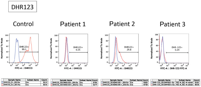
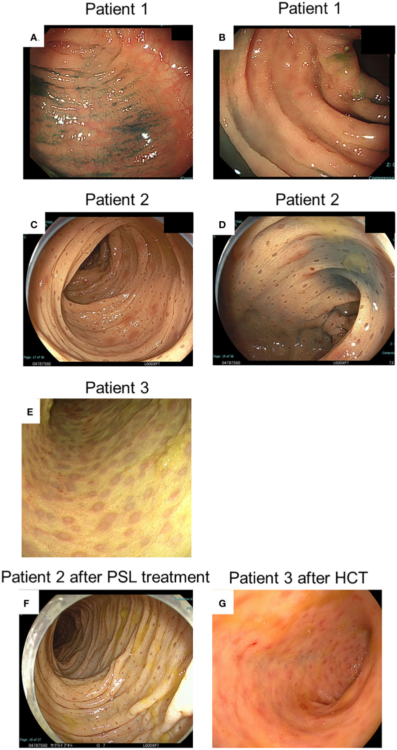
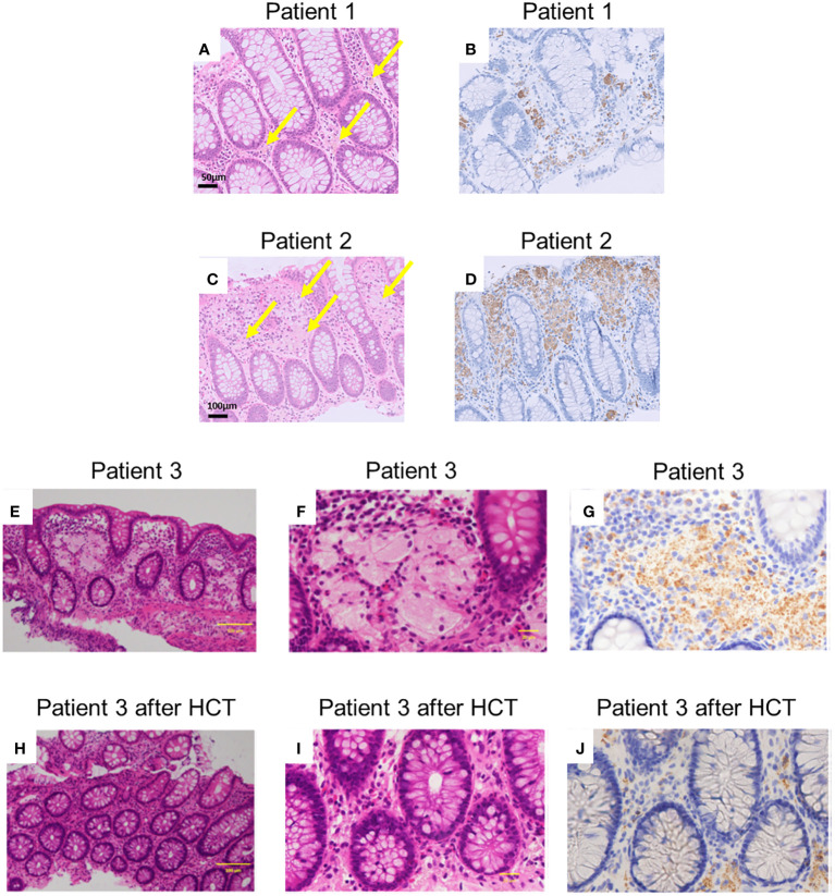
Background: Chronic granulomatous disease (CGD) is an inborn immune disorder in which the phagocytic system cannot eradicate pathogens, and autoinflammation occurs. Approximately half of the patients have associated gastrointestinal symptoms. Although most cases with CGD-associated colitis present nonspecific histology, colonoscopy in some cases shows brownish dots over a yellowish oedematous mucosa, which is termed a "leopard sign". However, the significance of these signs remains unclear.
Methods: We collected data from patients with CGD whose colonoscopic findings showed the leopard sign.
Results: Three patients with CGD and leopard signs were enrolled in this study. One patient underwent colonoscopy for frequent diarrhoea and weight gain failure, and another for anal fistula. The third patient was without gastrointestinal symptoms and underwent colonoscopy as a screening test before allogeneic haematopoietic cell transplantation (HCT). Endoscopic findings showed a mild leopard sign in the first case; however, non-contiguous and diffuse aphthae were observed throughout the colon. The other two cases were unremarkable except for the leopard sign. All the patients achieved remission with oral prednisolone or HCT. One patient underwent colonoscopy after HCT; results revealed improvements in endoscopy (including the leopard sign) and histological findings. However, another patient underwent colonoscopy after prednisolone treatment; this revealed no change in the leopard sign.
Conclusion: The leopard sign in the colon may be a characteristic endoscopic finding of CGD, even in patients who do not develop severe gastrointestinal symptoms; however, it does not reflect the severity of CGD-associated colitis.
Keywords: chronic granulomatous disease; colitis; endoscopy; haematopoietic cell transplantation; leopard sign.
Copyright © 2023 Nishikawa, Tomoda, Nakamura, Nagahama, Tanaka, Kanmura, Kirishima, Tanimoto, Okano, Kamiya, Okamoto, Kirimura, Morio, Okamoto and Kanegane.
Conflict of interest statement
The authors declare that the research was conducted in the absence of any commercial or financial relationships that could be construed as a potential conflict of interest.
Figures
Similar articles
-
Leopard Skin-Like Colonic Mucosa: A Novel Endoscopic Finding of Chronic Granulomatous Disease-Associated Colitis.J Pediatr Gastroenterol Nutr. 2016 Jan;62(1):56-9. doi: 10.1097/MPG.0000000000000905. J Pediatr Gastroenterol Nutr. 2016. PMID: 26164846
-
Chronic Granulomatous Disorder-Associated Colitis Can Be Accurately Evaluated with MRI Scans and Fecal Calprotectin Level.J Clin Immunol. 2019 Jul;39(5):494-504. doi: 10.1007/s10875-019-00651-2. Epub 2019 Jun 6. J Clin Immunol. 2019. PMID: 31172380 Free PMC article.
-
Intractable colitis associated with chronic granulomatous disease in a young girl.Turk J Pediatr. 2015 Mar-Apr;57(2):189-91. Turk J Pediatr. 2015. PMID: 26690604
-
Advances in the diagnosis and treatment of chronic granulomatous disease.Curr Opin Hematol. 2011 Jan;18(1):36-41. doi: 10.1097/MOH.0b013e32834115e7. Curr Opin Hematol. 2011. PMID: 21076296 Review.
-
Recent advances in chronic granulomatous disease.J Infect. 2014 Nov;69 Suppl 1:S32-5. doi: 10.1016/j.jinf.2014.07.013. Epub 2014 Sep 26. J Infect. 2014. PMID: 25264161 Review.
References
Publication types
MeSH terms
Substances
LinkOut - more resources
Full Text Sources