. 2023 Jun 5;47(4):247-261.
doi: 10.55730/1300-0152.2660. eCollection 2023.

Soloxolone methyl induces apoptosis and oxidative/ER stress in breast cancer cells and target cancer stem cell population

Affiliations

Soloxolone methyl induces apoptosis and oxidative/ER stress in breast cancer cells and target cancer stem cell population

Elif Ertürk et al. Turk J Biol. .

Abstract

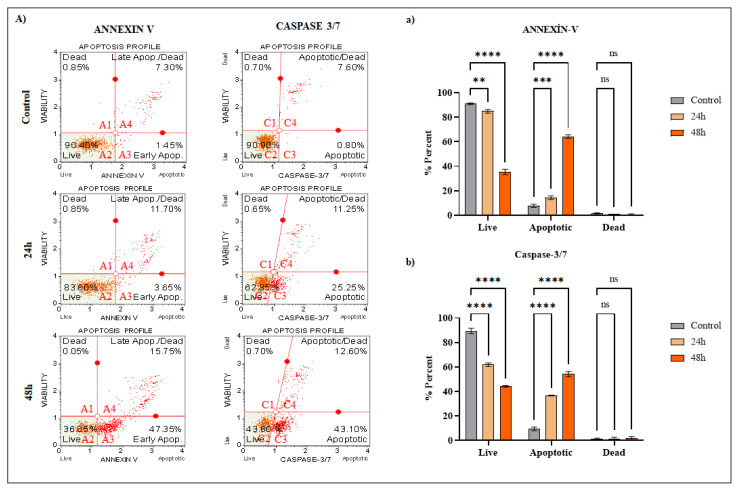
One of the most prevalent malignancies in women and one of the leading causes of cancer-related death is breast cancer. There is a need for new treatment approaches and drugs for breast cancer. Many studies show the high potential of triterpene compounds and their semisynthetic derivatives as anticancer agents due to their ability to induce apoptosis and suppress tumorigenesis. The effects of soloxolone methyl (SM), a semisynthetic derivative of 18-H-glycyrrhetinic acid, on the cytotoxicity and apoptosis of human breast cancer cell line (T-47D) and cancer stem cell (CSCs) population (mammospheres; CD44+/CD24-antigen) derived from breast cancer cells, were examined in this work. The ATP assay was used to determine SM growth-inhibitory effects. Fluorescent staining, caspase-cleaved cytokeratin 18, and flow cytometry analysis were used to determine the mode of the cell death. In addition, cell death was investigated at protein and gene levels by Western Blotting and PCR, respectively. SM resulted in cytotoxicity in a time and dose dependent manner via ROS production and ER stress in T-47D cells in 2 models. The mode of cell death was apoptosis, evidenced by phosphatidylserine exposure, caspase activation, and bax overexpression. In mammospheres as 3D model, SM decreased stem cell properties and induced cell death. Taken together, SM may be a promising agent in the treatment of breast cancer, especially due to its antigrowth activity on CSCs.

Keywords: Mammosphere; apoptosis; breast cancer; soloxolone methyl.


Conflict of interest statement

Declaration of competing interest: The authors declare that they have no known competing financial interests or personal relationships that could have appeared to influence the work reported in this paper.

Figures

Figure 1
Figure 1
(A) Chemical structure of 18βH-Glycyrrhetinic acids (a) and SM (b) (Markov et al., 2019). (B) Viability of T-47D cell treated with SM for 24 and 48 h. ATP assays were performed as cell viability assays. *Indicates statistically significant differences compared to untreated control: ***(p < 0.001). Data are presented as mean ± SD (n = 3).
Figure 2
Figure 2
(A) Phase/contrast and fluorescence microscopes images of T-47D breast cancer cells stained with Hoechst 33342 (blue) and Propidium iodide (red) and treated with 10 μM SM for 24 and 48 h. Data were presented as mean ± SD (n = 2). (B) Detection of M30-antigen levels after 10 μM dose of treatment with SM for 24 and 48 h. #Denotes statistically significant differences between groups (Control and 10 μM SM): *** (p < 0.001), # no significant. Data are presented as mean ± SD (n = 3). (C) Flow cytometry of T-47D cells treated with 10 μM SM for 24 h and 48 h: Analysis results of oxidative stress assay. (D) Viability analysis results of T-47D cells inhibited by NAC and treated with SM for 48 h.
Figure 3
Figure 3
(A) Flow cytometry of T-47D cells treated with 10 μM SM for 24 and 48 h: Analysis results of annexin V and caspase 3/7. (a) Gradhpad statistical results of annexin V flow cytometry analysis. (b) Gradhpad statistical results of caspase 3/7 flow cytometry analysis. *Indicates statistically significant differences compared to untreated control: ****(p < 0.001). ns; indicates statistically insignificant differences compared to the untreated control. Data are presented as mean ± SD (n = 3).
Figure 4
Figure 4
(A) Examination of apoptosis and ER-stress markers to elucidate apoptosis pathway. With the use of the ImageJ program, densitometry was carried out. GAPDH, or glyceraldehyde 3-phosphate dehydrogenase, was used as a reference point for quantification of the detected bands’ intensity. (B) Changes in the gene expression profiles of T-47D, determined by the simultaneous PCR method, after 48 h of treatment of SM compound with IC70 doses determined according to the ATP method. *Denotes statistically significant differences in comparison with control: *(p < 0.05), **(p < 0.01). Data are presented as mean ± SD (n=3). GAPDH, glyceraldehyde 3-phosphate dehydrogenase; RT-qPCR, quantitative real-time polymerase chain reaction; SD, standard deviation.
Figure 5
Figure 5
(A) Flow Cytometry results of CD44 and CD24 cells ratios of T-47D human breast cancer stem cell-like cells. (B) Phase-contrast microscope photographs of mammosphere cells after 48 h of 3.12–50 μM doses of SM treatment. (C) Viability of mammosphere cells treated with 3.12–50 μM doses of SM for 48 h. ATP assay were performed as cell viability assays. *Indicates statistically significant differences compared to untreated control: **(p < 0.01), ***(p < 0.001). Data are presented as mean ± SD (n = 3).
Figure 6
Figure 6
Fluorescence microscopes images of BCSCs stained with Hoechst 33342 (blue) and Propidium iodide (red) and treated with 50–12.5 μM doses of SM for 24 h. Data were presented as mean ± SD (n = 2).
Figure 7
Figure 7
(A) Examination of apoptosis and ER-stress markers to elucidate apoptosis pathway. With the use of the ImageJ program, densitometry was carried out. GAPDH, or glyceraldehyde 3-phosphate dehydrogenase, was used as a reference point for quantification of the detected bands’ intensity. (B) Changes in the gene expression profiles of BCSCs, determined by the simultaneous PCR method, after 48 h of treatment of SM compound with IC70 doses determined according to the ATP method. *Denotes statistically significant differences in comparison with control: *(p < 0.05), **(p < 0.01). Data are presented as mean ± SD (n = 3). GAPDH, glyceraldehyde 3-phosphate dehydrogenase; RT-qPCR, quantitative real-time polymerase chain reaction; SD, standard deviation.

References

    1. Akgun O, Erkisa M, Ari F. Effective and new potent drug combination: histone deacetylase and Wnt/β-catenin pathway inhibitors in lung carcinoma cells. Journal of Cellular Biochemistry. 2019;120(9):15467–15482. doi: 10.1002/jcb.28813. - DOI - PubMed
    1. Al-Hajj M, Wicha MS, Benito-Hernandez A, Morrison SJ, Clarke MF. Prospective identification of tumorigenic breast cancer cells. Proceedings of the National Academy of Sciences. 2003;100(7):3983–3988. doi: 10.1073/pnas.0530291100. - DOI - PMC - PubMed
    1. Alper P, Erkisa M, Genckal HM, Sahin S, Ulukaya E, et al. Synthesis, characterization, anticancer and antioxidant activity of new nickel (II) and copper (II) flavonoid complexes. Journal of Molecular Structure. 2019;1196:783–792. doi: 10.1016/j.molstruc.2019.07.009. - DOI
    1. Alper P, Salomatina OV, Salakhutdinov NF, Ulukaya E, Ari F. Soloxolone methyl, as a 18βH-glycyrrhetinic acid derivate, may result in endoplasmic reticulum stress to induce apoptosis in breast cancer cells. Bioorganic & Medicinal Chemistry . 2021;30:115963. doi: 10.1016/j.bmc.2020.115963. - DOI - PubMed
    1. Ames BN, Gold LS, Willett WC. The causes and prevention of cancer. Proceedings of the National Academy of Sciences. 1995;92(12):5258–5265. doi: 10.1073/pnas.92.12.5258. - DOI - PMC - PubMed

LinkOut - more resources