Ethyl acetate extract of Terminalia chebula alleviates DSS-induced ulcerative colitis in C57BL/6 mice
- PMID: 38152693
- PMCID: PMC10751924
- DOI: 10.3389/fphar.2023.1229772
Ethyl acetate extract of Terminalia chebula alleviates DSS-induced ulcerative colitis in C57BL/6 mice
Abstract
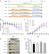
Background: The Chinese pharmacopeia records Terminalia chebula as effective in treating prolonged diarrhea and dysentery, blood in the stool, and prolapse. Modern pharmacological research proves it has multiple pharmacological benefits, including antioxidant, anti-inflammatory, analgesic, hepatoprotective, neuroprotective, and other properties. Objectives: This study aims to clarify the role of Terminalia chebula's ethyl acetate extract (TCEA) on ulcerative colitis (UC) induced by dextran sodium sulfate (DSS) in mice, as well as explore the potential mechanism of action. Materials and methods: The variation of different extracts of T. chebula was detected using the HPLC technique, and the main components in TCEA were identified. DSS was used to establish a mouse model to mimic the physiological state of UC in humans; the alleviating effect of TCEA and positive control 5-ASA on UC mice were evaluated by gavage treatment. Disease progression was assessed by monitoring the mouse's weight change and disease activity index (DAI). The changes in colon tissue were estimated by measuring colon length, HE, and AB-PAS staining and detecting oxidative stress parameters. The results draw from Western blot and real-time PCR showed the TLR4/MyD88/NF-κB pathway may involve in the anti-inflammatory activity of TCEA. Furthermore, the gut flora sequencing technique was employed to monitor the differentiation of intestinal microbiota of mice induced by DSS and TCEA treatment. Results: TCEA significantly lowered DAI scores and inhibited the weight loss and colonic shortening induced by DSS. The colon histomorphology and oxidative stress levels were enhanced after TCEA treatment compared with DSS induced UC group. TCEA attenuated the inflammatory response by regulating TLR4/MyD88/NF-κB pathway activation. Intestinal flora sequencing showed that DSS and TCEA greatly impacted mice's composition and diversity of intestinal microorganisms. But TCEA increased the abundance of Bacteroidetes and decreased the abundance of Firmicutes and Proteobacteria compared with the DSS group, which contributed a lot to returning the intestinal flora to a balanced state. Conclusion: This study confirms the alleviating effect of TCEA on UC and provides new ideas for developing TCEA into a new drug to treat UC.
Keywords: TLR4/MyD88/NF-κB; Terminalia chebula; inflammation; intestinal flora; ulcerative colitis.
Copyright © 2023 Dong, Li, Liu, Zhou and Chen.
Conflict of interest statement
The authors declare that the research was conducted in the absence of any commercial or financial relationships that could be construed as a potential conflict of interest.
Figures


Similar articles
-
Sanhuang Xiexin decoction ameliorates DSS-induced colitis in mice by regulating intestinal inflammation, intestinal barrier, and intestinal flora.J Ethnopharmacol. 2022 Oct 28;297:115537. doi: 10.1016/j.jep.2022.115537. Epub 2022 Jul 14. J Ethnopharmacol. 2022. PMID: 35843414
-
Investigating the mechanism of enhanced medicinal effects of Terminalia chebula fruit after processing based on intestinal flora and metabolomics.Int Immunopharmacol. 2024 Dec 25;143(Pt 1):113271. doi: 10.1016/j.intimp.2024.113271. Epub 2024 Oct 4. Int Immunopharmacol. 2024. PMID: 39368133
-
Ganluyin ameliorates DSS-induced ulcerative colitis by inhibiting the enteric-origin LPS/TLR4/NF-κB pathway.J Ethnopharmacol. 2022 May 10;289:115001. doi: 10.1016/j.jep.2022.115001. Epub 2022 Jan 24. J Ethnopharmacol. 2022. PMID: 35085745
-
A comprehensive review on the diverse pharmacological perspectives of Terminalia chebula Retz.Heliyon. 2022 Aug 14;8(8):e10220. doi: 10.1016/j.heliyon.2022.e10220. eCollection 2022 Aug. Heliyon. 2022. PMID: 36051270 Free PMC article. Review.
-
The potential of Terminalia chebula in alleviating mild cognitive impairment: a review.Front Pharmacol. 2024 Oct 18;15:1484040. doi: 10.3389/fphar.2024.1484040. eCollection 2024. Front Pharmacol. 2024. PMID: 39494343 Free PMC article. Review.
Cited by
-
Effects of Dietary Terminalia chebula Extract on Growth Performance, Immune Function, Antioxidant Capacity, and Intestinal Health of Broilers.Animals (Basel). 2024 Feb 28;14(5):746. doi: 10.3390/ani14050746. Animals (Basel). 2024. PMID: 38473130 Free PMC article.
-
Bacillus clausii spores maintain gut homeostasis in murine ulcerative colitis via modulating microbiota, apoptosis, and the TXNIP/NLRP3 inflammasome cascade.Toxicol Rep. 2024 Dec 16;14:101858. doi: 10.1016/j.toxrep.2024.101858. eCollection 2025 Jun. Toxicol Rep. 2024. PMID: 39802600 Free PMC article.
-
Huanglian Ejiao Decoction Alleviates Ulcerative Colitis in Mice Through Regulating the Gut Microbiota and Inhibiting the Ratio of Th1 and Th2 Cells.Drug Des Devel Ther. 2025 Jan 17;19:303-324. doi: 10.2147/DDDT.S468608. eCollection 2025. Drug Des Devel Ther. 2025. PMID: 39845151 Free PMC article.
-
Comprehensive Review on Fruit of Terminalia chebula: Traditional Uses, Phytochemistry, Pharmacology, Toxicity, and Pharmacokinetics.Molecules. 2024 Nov 24;29(23):5547. doi: 10.3390/molecules29235547. Molecules. 2024. PMID: 39683707 Free PMC article. Review.
-
Terminalia Chebula Extract Replacing Zinc Oxide Enhances Antioxidant and Anti-Inflammatory Capabilities, Improves Growth Performance, and Promotes Intestinal Health in Weaned Piglets.Antioxidants (Basel). 2024 Sep 5;13(9):1087. doi: 10.3390/antiox13091087. Antioxidants (Basel). 2024. PMID: 39334746 Free PMC article.
References
LinkOut - more resources
Full Text Sources

