. 2024 Jun 14;44(7):671-692.
doi: 10.1093/asj/sjad382.

The Ponytail Lift: 22 Years of Experience in 600 Cases of Endoscopic Deep Plane Facial Rejuvenation

The Ponytail Lift: 22 Years of Experience in 600 Cases of Endoscopic Deep Plane Facial Rejuvenation

Chia Chi Kao et al. Aesthet Surg J. .

Abstract

Background: During aging, the face loses volume with progressive sagging of the soft tissues, while the neck demonstrates skin laxity and muscle banding. The treatment of facial and neck aging usually involves a traditional facelift, which can cause noticeable scarring and distortion of anatomy.

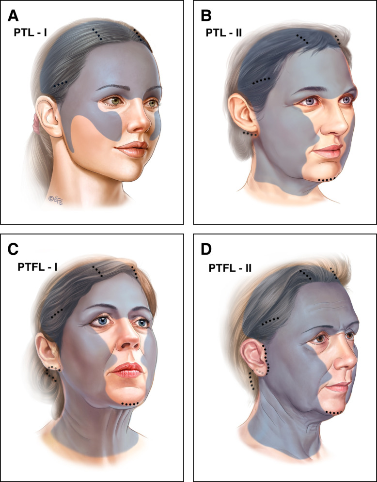
Objectives: Modern facelift surgery must avoid such shortcomings and still address aging in all layers of the face. To achieve this goal a novel surgical technique was developed and coined the "ponytail lift" (PTL). When global facial rejuvenation is indicated, this procedure is combined with neck skin excision and referred to as the "ponytail facelift" (PTFL).

Methods: A retrospective analysis of 600 consecutive cases over 22 years (2000-2022) of facial rejuvenation employing the endoscopic techniques of PTL and PTFL was performed. Patients were followed for at least 12 months postoperatively. Demographics, surgical data, and complications were recorded and analyzed. Additionally, technical details of the PTL and PTFL are discussed.

Results: There were no instances of postoperative skin flap necrosis, and no permanent nerve injuries were recorded. An additional surgical touch-up procedure to address unsatisfied aesthetic needs was performed in 20 cases.

Conclusions: The ponytail procedures offer a stepwise approach matched to the extent of the problem and are intended to refresh or transform the face with minimal incisions. The procedures represent a deep plane facelift without the scar burden, with incisions that are hidden in the temple, postauricular, and posterior scalp. The described techniques are safe and effective while providing reliable and satisfying results.

背景: 随着年龄的增长,面部体积会逐渐减小,软组织会逐渐下垂,而颈部会出现皮肤松弛和肌肉带状。面部和颈部衰老的治疗通常涉及传统的面部拉皮术,这会导致明显的疤痕和解剖结构扭曲。

目标: 现代面部拉皮手术必须避免此类缺点,同时还要解决面部所有层面的衰老问题。为了实现这一目标,开发了一种新颖的手术技术,创造了“马尾辫提升术”(PTL)这一名称。当需要进行整体面部年轻化时,该手术与颈部皮肤切除术相结合,称为“马尾辫拉皮术”(PTFL)。

方法: 回顾性分析了 22 年间(2000-2022 年)采用 PTL 和 PTFL 内窥镜技术进行面部年轻化的 600 个连续病例。术后对患者进行至少 12 个月的随访。记录并分析了人口统计学、手术数据和并发症。此外,还讨论了 PTL 和 PTFL 的技术细节。

结果: 没有出现术后皮瓣坏死的情况,也没有记录到永久性神经损伤。在 20 个病例中进行了额外的外科修补手术,以解决未满足的美学需求。

结论: 马尾辫手术提供了一种与问题程度相匹配的分步方法,旨在以最小的切口使面部得以改造或焕然一新。这些手术代表了一种深层平面拉皮术,没有疤痕负担,切口隐藏在太阳穴、耳后和头皮后部。所描述的技术是安全有效的,同时提供可靠且令人满意的结果。

证据等级:: 3 级(治疗性)

Level of Evidence: 3: formula image


Figures

Figure 1.
Figure 1.
The 4 ponytail types, the incisions, and the extent of the dissection. There are 4 types of ponytail procedures: (A) Ponytail Lift I (PTL-I) is designed for patients presenting in their 30s or 40s with signs of early aging, commonly seen in the upper two-thirds of the face, or younger patients for facial beautification. The incisions are all hidden in the scalp. (B) Ponytail Lift II (PTL-II) is typically indicated for patients 40 to 45 years of age with early jowling but only minimal skin laxity of the neck. A 2-cm endoscopic access incision in the postauricular sulcus is added to thin the lower face and jowls and allow endoscopic deep plane dissection and tightening of the SMAS layer. When submental fullness (double chin) is present, deep contouring of the anterior neck is added through a 1.5-cm incision under the chin, however, no skin in the neck is removed. (C) Ponytail Facelift I (PTFL-I) is indicated when there is enough laxity and redundancy of the lower face and neck skin requiring resection. The PTFL-I procedure includes an extended posterior auricular incision for a deep plane lower face and necklift. Correction of the anterior neck by deep contouring and platysma plication as well as application of a posterior corset is necessary. The typical patient age for this cohort is 45 to 65 years, presenting with jowls and a mild to moderate amount of neck skin laxity. Importantly, there are no incisions in the pretragal area or temporal hairline. (D) For a Ponytail Facelift II (PTFL-II) the typical age is 65 years and older. The issues to address include heavy jowling, significant skin excess in the neck, and poor skin tone and elasticity. A limited pretragal incision is added to the PTFL-I approach to help manage the significant excess skin. Importantly there is no incision in the sideburns or any extension into the temporal hairline. SMAS, superficial musculoaponeurotic system.
Figure 2.
Figure 2.
Surgical marking. Surgical marking includes the bony architecture, Pitanguy's line, the anticipated location of the sentinel vein in the temporal area 1 cm superior to the superior zygomatic arch and 1 cm lateral to the zygomatic process, and areas of desired fat transfer. For PTFL cases we mark the anterior border of the masseter muscle as well as a line from the earlobe to the ala of the nose delineating the upper limit of the subcutaneous lower face and neck dissection. PTFL, ponytail facelift.
Figure 3.
Figure 3.
Limited incision of lower face and neck lift. On the left (A), the dissection plane is immediately superficial to the platysma and the posterior neck fascia down to the clavicle, extending anteriorly across the midline of the neck and posteriorly to the anterior border of the trapezius muscle. The subcutaneous fat is lifted with the skin to preserve cutaneous blood supply. On the right (B), the dissection plane is kept immediately above the SMAS, preserving the subcutaneous fat and blood supply with the skin. The medial extent of the subcutaneous dissection approaches the oral commissure. It is also important to release the mandibular retaining ligaments. Any thickening of the subcutaneous fat in the jowl area is directly trimmed with scissors to ensure even thickness. SMAS, superficial musculoaponeurotic system.
Figure 4.
Figure 4.
Lower face and neck deep plane dissection. (A) The lateral edge of the platysma/SMAS is sharply dissected off of the parotid fascia. Medially, the dissection of the SMAS is extended 2 cm past the anterior boarder of the masseter muscle (fixed SMAS), into the mobile SMAS. Inferiorly, the platysma/SMAS dissection continues 3 to 4 cm below the jawline. The subplatysmal dissection in the neck is extended about 4 cm medially to the area of the submandibular glands. (B) The buccal branch of the facial nerve may be visualized running in the parotid masseteric fascia. Care is taken to avoid injury to the marginal mandibular branch of the facial nerve. (C) A 2.5-cm submental curvilinear keyhole incision is made very anteriorly on the chin pad skin. The plane of dissection is immediately above the platysma. At the level of the submental incision, a horizontal entry is made perpendicular to the platysmal muscle fibers to dissect and lift the platysma muscle. Subplatysmal fat is then resected. The anterior belly of the digastric muscle is debulked with needle cautery as needed for contour and then plicated with 4.0 Ethibond interrupted sutures. Submandibular gland reduction is performed when indicated. The anterior edge of the platysma is plicated with interrupted suture, starting 2 cm below the cricoid cartilage and proceeding all the way up to the chin incision to avoid a cobra neck deformity. A 2-cm back cut on the bands is made below the most inferior plication suture. SMAS, superficial musculoaponeurotic system.
Figure 5.
Figure 5.
Endoscopic access and dissection planes: total deep plane dissection from cranium to clavicle. The dissection is performed between deep and superficial temporal fascia and done completely endoscopically. In the forehead and brow region, dissection is performed in the subperiosteal plane, all the way down past the orbital rim. In the temporal area, dissection proceeds endoscopically between the superficial and deep temporal fascia and continues down toward the zygomatic arch. The sentinel vein and the medial and lateral zygomaticotemporal neurovascular bundles are identified and preserved. Dissection continues to the arch, and the arch ligament is sharply released. The zygomaticomalar ligament also needs to be released to gain access to the midface. In the midface, the dissection plane is between the SOOF and the orbicularis oculi muscle. The dissection continues along the zygomaticus major and minor muscles, down across the nasolabial fold. The zygomatic branch of the facial nerve must be preserved in this region. To minimize the risk of nerve injury it is necessary to stay on the body of the zygoma and proceed medially, avoiding any lateral deviation. The midface flap that is mobilized across the nasolabial fold is suspended from several fixation points to the deep temporal fascia. SOOF, suborbicularis oculi fat.
Figure 6.
Figure 6.
Ligamentous release points. For a successful and long-lasting forehead lift and brow rotation, there are 5 anatomical entities that need to be completely released: the superior temporal septum; temporal lateral adhesion; zygomatic arch ligament; inferior temporal septum; lateral orbital thickening; and external canthal tendon (if lateral canthal tilt is desired).
Figure 7.
Figure 7.
Suture tying sequence. After completion of the anterior platysma corset the following suture placement algorithm is extremely important to maximize vertical rotational tissue displacement. (1) We begin with the superior zygomatic arch sutures to the temporal fascia, usually 2 to 3 laterally in the “safe lane.” The exact positioning is determined by the relationship of the medial and lateral zygomatic lateral neurovascular bundles. (2) Next, we place 2 or 3 cheek sutures lifting the cheek fat pads toward the temple area. (3, 4) The next level of suturing connects the bitemporal (anterior wound edge) and paramedian (posterior wound edge) incisions to perform the brow rotation. (5) As a next step we place stiches that grab the TPF island on its distal cut edge and tie them to the temporal fascia. (6) The proximal edge of the TPF island flap is sutured to the temporal incision wound edge and fascia. (7) Finally, the posterior platysma corset is completed. Importantly, the assistant lifts the face vertically during the entire suture tying sequence, with hooks deploying tension from the paramedian incisions. TPF, temporoparietal fascia.
Figure 8.
Figure 8.
Progressive tension concept of the vertical-central lift (Fabergé vector). Tying the sutures from bottom to top enables an ideal brow positioning and vertical lifting of all tissues from clavicle to cranium, as depicted in (A). This leads to a heart shaped tapered face with the ideal proportions of an inverse Fabergé egg, as depicted in (B).
Figure 9.
Figure 9.
Representative result of Ponytail Lift Type I. This 36-year-old female patient was battling early signs of aging (sagging of the brows, hooding of the upper lids, drooping of the cheeks, deepening of the nasolabial folds, formation of jowls). She had been receiving fillers for the last 8 years unsuccessfully trying to lift her face, resulting in distortion of her delicate features. We dissolved the fillers in her cheeks, undereye areas, and jawline, performed a Ponytail Lift Type I and an upper lip lift. This allowed us to restore the delicate features and harmonize the face without any visible scars. Parts A, C, E, and G show the patient preoperatively, and parts B, D, F, and H show her results 1 year postoperatively.
Figure 10.
Figure 10.
Representative result of Ponytail Lift Type II. This 42-year-old female patient desired facial rejuvenation because of laxity of the upper eyelids and flat cheeks with early jowling. She also complained about submental fullness, an undefined jawline, and neck laxity. However, skin quality overall was still good and there was not enough skin redundancy to require resection. Therefore, her needs were addressed with a Ponytail Lift Type II without any skin removal. Additionally, correction of an unsatisfactory lip lift scar was performed. (A), (C), (E), and (G) show the patient preoperatively, and (B), (D), (F), and (H) show her results [x] years postoperatively.
Figure 11.
Figure 11.
Representative result of Ponytail Facelift Type I. This 57-year-old female patient is now 2 years following her Ponytail Facelift Type I. She complained about general signs of aging with a particular focus on her neck. We lifted the entire deep plane from the top of her head to the bottom of her neck and tightened it without the traditional facelift incisions along the temple hairline and in front of the ear. An upper lip lift with an internal corner lift was also performed. (A), (C), (E), and (G) show the patient preoperatively, and (B), (D), (F), and (H) show her results 2 years postoperatively.
Figure 12.
Figure 12.
Representative result of Ponytail Facelift Type I with extended follow-up. This 69-year-old female patient received a Ponytail Facelift Type I 17 years ago. No fillers or other minimal invasive treatments or surgical interventions have been performed elsewhere in the meantime. Even after this period of time, her brow has maintained its corrected position and the midface and neck are still satisfactory. (A), (C), (E), (G), and (I) show the patient preoperatively, and (B), (D), (F), (H) and (J) show her results 17 years postoperatively.
Figure 13.
Figure 13.
Representative result of Ponytail Facelift Type II. We performed a Ponytail Facelift Type II in this 58-year-old female patient to achieve a panfacial rejuvenation from the top of the head to the bottom of the neck. She was looking for a transformation in the shape of her face: from a square jowly shape to a V-shape of youth. (A), (C), (E), and (G) show the patient preoperatively, and (B), (D), (F), and (H) show her results 3 years postoperatively.

Comment in

Similar articles

Cited by

References

    1. Swift A, Liew S, Weinkle S, Garcia JK, Silberberg MB. The facial aging process from the “inside out”. Aesthet Surg J. 2021;41(10):1107–1119. doi: 10.1093/asj/sjaa339 - DOI - PMC - PubMed
    1. Carniol PJ, Ganc DT. Is there an ideal facelift procedure? Curr Opin Otolaryngol Head Neck Surg. 2007;15(4):244–252. doi: 10.1097/MOO.0b013e32825a678d - DOI - PubMed
    1. Hashem AM, Couto RA, Surek C, Swanson M, Zins JE. Facelift part II: surgical techniques and complications. Aesthet Surg J. 2021;41(10):NP1276–NPNP94. doi: 10.1093/asj/sjab081 - DOI - PubMed
    1. Hashem AM, Couto RA, Duraes EFR, et al. . Facelift part I: history, anatomy, and clinical assessment. Aesthet Surg J. 2020;40(1):1–18. doi: 10.1093/asj/sjy326 - DOI - PubMed
    1. Luu NN, Friedman O. Facelift surgery: history, anatomy, and recent innovations. Facial Plast Surg. 2021;37(5):556–563. doi: 10.1055/s-0040-1715616 - DOI - PubMed