. 2024 Mar 12;149(11):860-884.
doi: 10.1161/CIRCULATIONAHA.123.065517. Epub 2023 Dec 28.

Metabolic Communication by SGLT2 Inhibition

Affiliations

Metabolic Communication by SGLT2 Inhibition

Anja M Billing et al. Circulation. .

Abstract

Background: SGLT2 (sodium-glucose cotransporter 2) inhibitors (SGLT2i) can protect the kidneys and heart, but the underlying mechanism remains poorly understood.

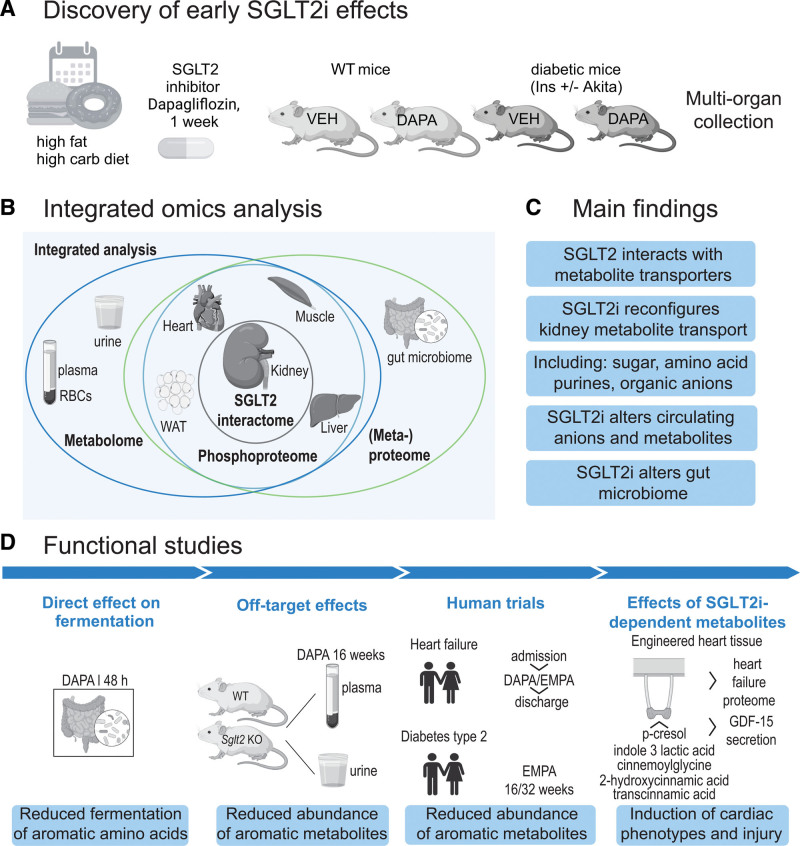
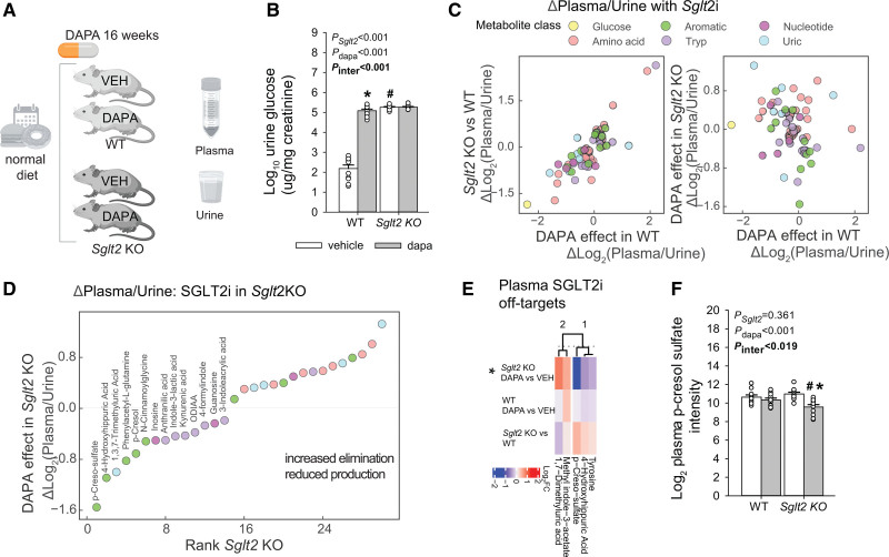
Methods: To gain insights on primary effects of SGLT2i that are not confounded by pathophysiologic processes or are secondary to improvement by SGLT2i, we performed an in-depth proteomics, phosphoproteomics, and metabolomics analysis by integrating signatures from multiple metabolic organs and body fluids after 1 week of SGLT2i treatment of nondiabetic as well as diabetic mice with early and uncomplicated hyperglycemia.

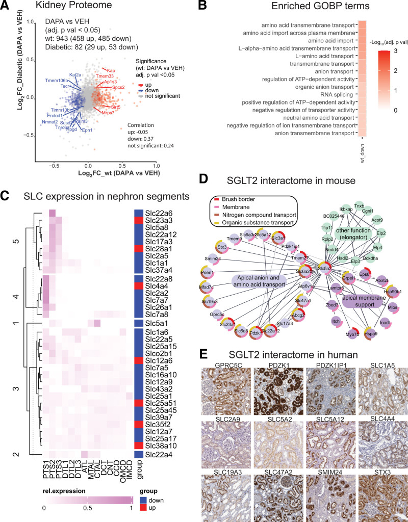
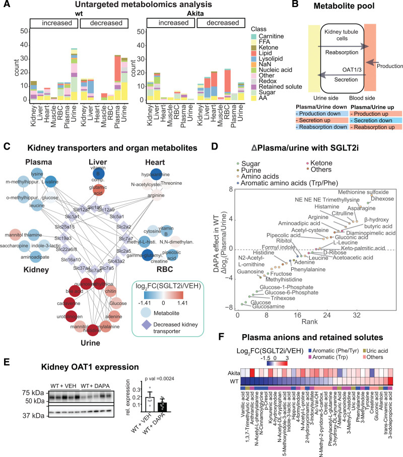
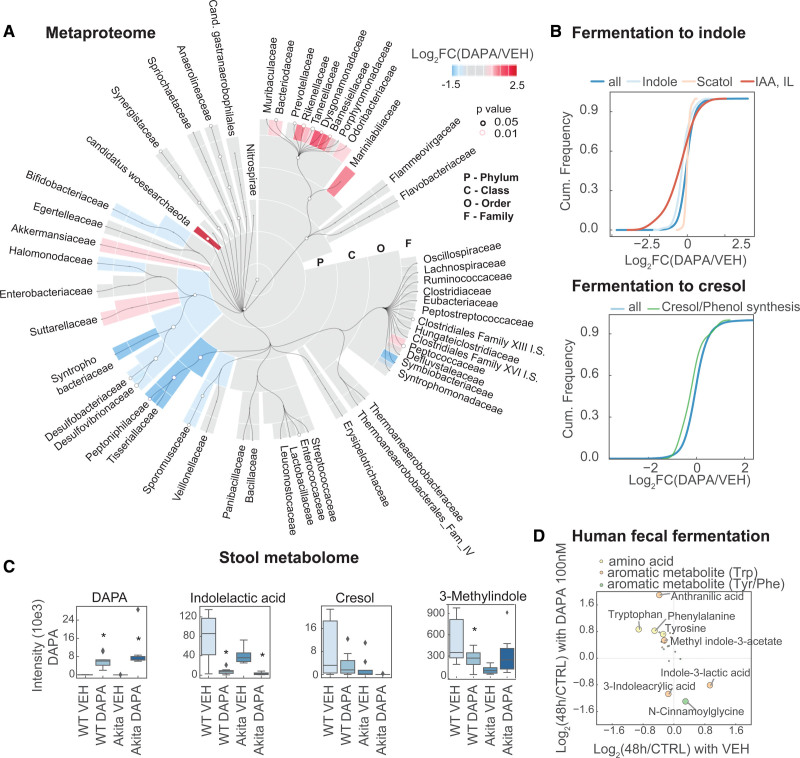
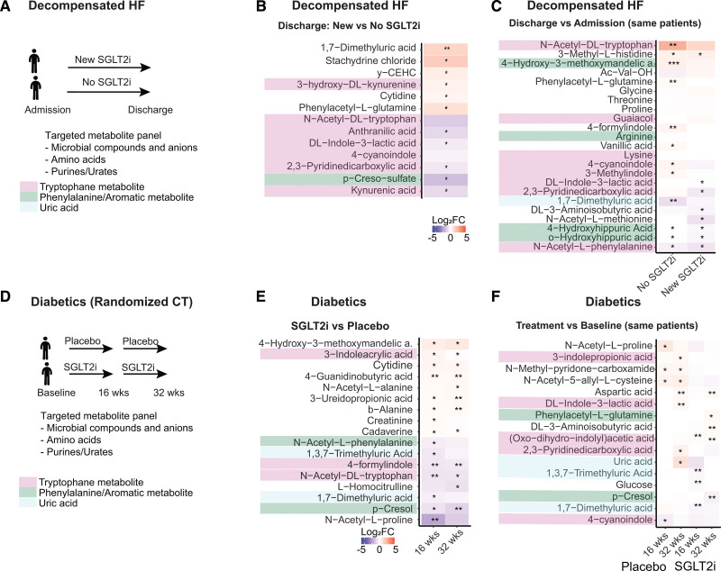
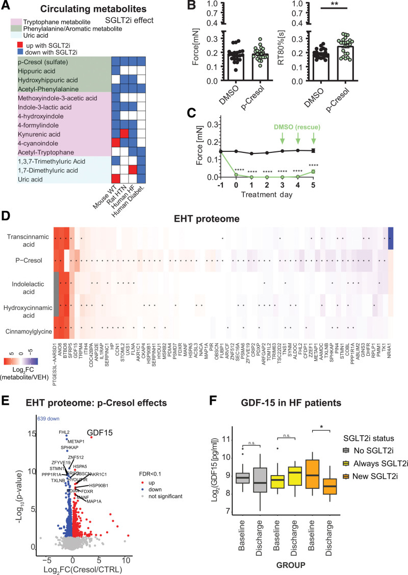
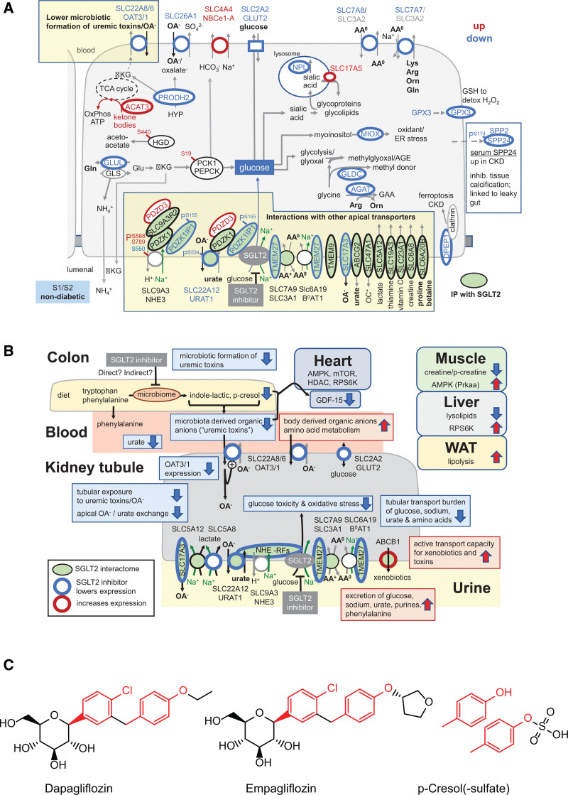
Results: Kidneys of nondiabetic mice reacted most strongly to SGLT2i in terms of proteomic reconfiguration, including evidence for less early proximal tubule glucotoxicity and a broad downregulation of the apical uptake transport machinery (including sodium, glucose, urate, purine bases, and amino acids), supported by mouse and human SGLT2 interactome studies. SGLT2i affected heart and liver signaling, but more reactive organs included the white adipose tissue, showing more lipolysis, and, particularly, the gut microbiome, with a lower relative abundance of bacteria taxa capable of fermenting phenylalanine and tryptophan to cardiovascular uremic toxins, resulting in lower plasma levels of these compounds (including p-cresol sulfate). SGLT2i was detectable in murine stool samples and its addition to human stool microbiota fermentation recapitulated some murine microbiome findings, suggesting direct inhibition of fermentation of aromatic amino acids and tryptophan. In mice lacking SGLT2 and in patients with decompensated heart failure or diabetes, the SGLT2i likewise reduced circulating p-cresol sulfate, and p-cresol impaired contractility and rhythm in human induced pluripotent stem cell-derived engineered heart tissue.

Conclusions: SGLT2i reduced microbiome formation of uremic toxins such as p-cresol sulfate and thereby their body exposure and need for renal detoxification, which, combined with direct kidney effects of SGLT2i, including less proximal tubule glucotoxicity and a broad downregulation of apical transporters (including sodium, amino acid, and urate uptake), provides a metabolic foundation for kidney and cardiovascular protection.

Keywords: diabetes mellitus; gastrointestinal microbiome; heart; kidney; metabolome; plasma; proteome; sodium-glucose transporter 2 inhibitors; uremic toxins; urine.


Conflict of interest statement

Disclosures Dr Rinschen declares pending research funding from Novo Nordisk unrelated to this work. Over the past 12 months, Dr Vallon has served as a consultant for Lexicon and received speaker honoraria from AstraZeneca and grant support for investigator-initiated research from AstraZeneca, Boehringer Ingelheim, Gilead, Lexicon, Maze, Merck, and Novo-Nordisk. Dr Magnussen receives study-specific funding from the German Center for Cardiovascular Research (DZHK; Promotion of Women Scientists Programme; FKZ 81X3710112), the Deutsche Stiftung für Herzforschung, the Dr Rolf M. Schwiete Stiftung, NDD, and Loewenstein Medical unrelated to the current work. Dr Magnussen has received speaker fees from AstraZeneca, Novartis, Boehringer Ingelheim/Lilly, Bayer, Pfizer, Sanofi, Aventis, Apontis, and Abbott outside this work. Dr Dugourd and R. Fallegger report funding from Pfizer. Dr Saez-Rodriguez reports funding from GSK, Pfizer, and Sanofi and fees from Travere Therapeutics, Stadapharm, and Astex. Dr Hoxha served on advisory boards for Novartis, Morphosys AG, Sotio, and Argenx. The other authors declare no conflict of interest.

Figures

Figure 1.
Figure 1.
Overview of study design and phenotypes. A, Overall discovery strategy. Wild-type (WT) and hyperglycemic type 1 diabetic Akita mice were put on a high-fat, high-carbohydrate Western diet for 4 weeks, followed by SGLT2i (sodium-glucose cotransporter 2 inhibitor), dapagliflozin (10 mg/kg of Western diet), or vehicle (repelleted Western diet) for 1 week. B, Organs and body fluids were snap-frozen and subjected to multi-omics analysis using mass spectrometry. SGLT2 interactome was determined from kidney. C, Main findings. D, Overview of functional follow-up studies and key findings. GDF15 indicates growth/differentiation factor 15; RBC, red blood cell; and WAT, white adipose tissue.
Figure 2.
Figure 2.
Integrated proteome/interactome analysis indicates broad remodeling of kidney metabolite transport by SGLT2i. A, Volcano plot quantification of proteomics analysis of kidney cortex. Overlapping significant proteins (false discovery rate <0.05) between wild-type (WT) and Akita mice upon SGLT2i (sodium-glucose cotransporter 2 inhibitor) treatment are labeled with their gene name. B, Enrichment of Gene Ontology Biological Process (GOBP) terms in WT kidney cortex (kidney cortex proteome was used as background). C, Nephron localization of altered transmembrane transporters of the solute carrier group (SLC). D, Mass spectrometry–based interactome analysis of mouse SGLT2i. SGLT2 interacts with membrane transporters that mediate the reabsorption or secretion of sodium, amino acids, urate, protons, anions, and multiple other compounds. E, Human tissue atlas of selected putative human SGLT2 interactors (source: The Human Proteome Atlas; https://www.proteinatlas.org).
Figure 3.
Figure 3.
Untargeted metabolome analysis reveals SGLT2i-induced alterations in amino acid and organic anion metabolism. A, Overview of untargeted metabolomics results in wild-type (WT) and diabetic Akita mice. Significant metabolites from >30 000 quantified features with breakdown of altered metabolites by organ and genotype. B, The metabolite pool is sensitive to changes in production as well as kidney secretion and reabsorption. C, Illustration of interorgan communication by linking solute carrier group (SLCs) downregulated in the kidney to enhanced urinary metabolites and depleted metabolites in other organs. D, SGLT2 (sodium-glucose cotransporter 2)–dependent changes in plasma:urine ratios indicate increased urinary excretion of glucose and glucose metabolites, and less secretion of substrates of the kidney organic anion transporters (see text for details). Urine metabolites were normalized to creatinine. E, Immunoblot analysis of OAT1 (organic anion transporter 1) in SGLT2i-treated WT mice shows reduction of OAT1 (Slc22a6; normalized to b-actin; 2-sided t test), consistent with proteomics results. F, Targeted analysis of microbiota-derived organic anions (uremic toxins) in the plasma of WT and diabetic Akita mice. RBC indicates red blood cell.
Figure 4.
Figure 4.
Metaproteomics analysis demonstrates reshaping and attenuation of the microbiome fermentation of amino acids to uremic toxins by SGLT2i. A, Taxonomic analysis of metaproteome of gut microbiota with SGLT2i (sodium-glucose cotransporter 2 inhibitor) on family level. B, Cumulative histogram analysis of cresol- and phenol-producing bacteria species on metaproteome level, as well as tryptophan-metabolizing bacteria. C, Murine fecal metabolomics results of key metabolites. D, Targeted metabolomics of human fecal microbiota fermentation (48 hours) in the presence or absence of SGLT2i dapagliflozin (100 nM). Metabolites with >0.5-fold change (48 hours fermentation versus control) and P<0.05 between both conditions are labeled with an open circle (3 donors, with 3 fermentations each). IAA indicates indoleacrylic acid; and IL, indolelactate.
Figure 5.
Figure 5.
Using Sglt2 KO mice to probe for metabolic off-target effects of SGLT2i. A, Study design. B, Quantification of glucosuria. C, Quantification of plasma:urine ratios in dapagliflozin-treated mice and Sglt2 knockout (KO) mice as compared with untreated wild-type (WT) mice. D, Rank of changes in plasma:urine ratios with SGLT2i (sodium-glucose cotransporter 2 inhibitor; all P<0.05). E, Analysis of off-target effects on mouse plasma, defined as significant changes in the presence of SGLT2i in Sglt2 KO mice. F, Quantification of p-cresol sulfate abundance in response to SGLT2i in both WT and Sglt2 KO mice. B and F, 2-way ANOVA to probe for a significant effect of Sglt2 genotype (Sglt2), dapagliflozin (dapa), or the interaction between the 2 factors (Pinteraction). If the interaction was statistically significant, then a pairwise multiple comparison procedure (Holm-Sidak method) identified the significant effects. *P<0.05 versus vehicle; #P<0.05 versus WT.
Figure 6.
Figure 6.
SGLT2i-induced reduction in uremic toxins translates to humans. A and D, Design of decompensated heart failure (HF) cohort and randomized controlled trial in patients with diabetes. B and C, Decompensated HF: heatmaps of SGLT2i (sodium-glucose cotransporter 2 inhibitor)–induced log2 fold changes for comparisons at discharge (B) and between admission and discharge (C). D, Randomized control trial of patients with diabetes: heatmaps of SGLT2i-induced log2 fold changes for SGLT2i versus placebo (E) and treatment versus baseline (F). SGLT2i treatment reduces uremic toxins originating from aromatic amino acid fermentation.
Figure 7.
Figure 7.
Effects of SGLT2i-modulated uremic toxin, p-cresol, on human engineered heart tissue. A, Summary of metabolite changes in response to SGLT2i (sodium-glucose cotransporter 2 inhibitor) in mouse, rat, and human. B, Human engineered heart tissue (EHT) was exposed to either DMSO (black) or 300 µM p-cresol (PCL; green) for 5 days. Bar charts show force (mN), and relaxation time from peak to 80% relaxation (RT 80%; seconds) in spontaneously contracting EHTs. C, Time course of spontaneous force 1 day before exposure to 3 mM PCL (−1; green) or DMSO (black) until 5 days of daily treatment. After 3 days of treatment, PCL was replaced by DMSO (green arrows) for another 48 hours’ incubation (n=16–22 per group, derived from 4 [300 µM] and 5 [3 mM] independent rounds of cardiac differentiation unpaired, parametric T test [** = P<0.01 and **** = P<0.0001]). D and E, Proteomic effects of incubation of EHT with p-cresol (300 µM) or other SGLT2i-modulated metabolites for 5 days. F, GDF15 (growth/differentiation factor 15) in patients with heart failure (HF) with new SGLT2i treatment (patients from Figure 6A). FDR indicates false discovery rate; HTN, hypertension; and WT, wild type.
Figure 8.
Figure 8.
Integrated views on the reconfiguration by SGLT2i of the early proximal tubule and systemic metabolic communication. A, Summary of findings and metabolic communication of SGLT2i (sodium-glucose cotransporter 2 inhibitor) in an early proximal tubule centric view in wild-type (WT) mice. Proteins with red lining were upregulated and proteins with blue lining were downregulated by SGLT2i. Proteins showing green filling have been shown to interact with SGLT2 in affinity purification analysis (see also Figure 2D). For further discussion, see text and Figure S8. B, Summary of findings and metabolic communication of SGLT2i on a systemic level in WT mice. C, Chemical structures of SGLT2 inhibitors dapagliflozin and empagliflozin and p-cresol. ACAT3 indicates acetyl-coenzyme A acetyltransferase 3; AGE, advanced glycation end products; AMPK, 5′-adenosine monophosphate–activated protein kinase; CKD, chronic kidney disease; ER, endoplasmic reticulum; GLUT2, glucose transporter 2; HGD, homogentisate 1,2-dioxygenase; NHE3, Na+/H+ exchanger 3; OAT1, organic anion transporter 1; PDZK1IP1, PDZK1 interacting protein 1; SLC, solute carrier; TMEM27, transmembrane protein 27; and WAT, white adipose tissue.

Comment in

References

    1. Vallon V. Glucose transporters in the kidney in health and disease. Pflugers Arch. 2020;472:1345–1370. doi: 10.1007/s00424-020-02361-w - PMC - PubMed
    1. Herrington WG, Staplin N, Wanner C, Green JB, Hauske SJ, Emberson JR, Preiss D, Judge P, Mayne KJ, Ng SYA, et al. ; The EMPA-KIDNEY Collaborative Group. Empagliflozin in patients with chronic kidney disease. N Engl J Med. 2023;388:117–127. doi: 10.1056/NEJMoa2204233 - PMC - PubMed
    1. Anker SD, Butler J, Filippatos G, Ferreira JP, Bocchi E, Böhm M, Brunner-La Rocca H-P, Choi D-J, Chopra V, Chuquiure-Valenzuela E, et al. ; EMPEROR-Preserved Trial Investigators. Empagliflozin in heart failure with a preserved ejection fraction. N Engl J Med. 2021;385:1451–1461. doi: 10.1056/NEJMoa2107038 - PubMed
    1. Heerspink HJL, Stefánsson BV, Correa-Rotter R, Chertow GM, Greene T, Hou F-F, Mann JFE, McMurray JJV, Lindberg M, Rossing P, et al. ; DAPA-CKD Trial Committees and Investigators. Dapagliflozin in patients with chronic kidney disease. N Engl J Med. 2020;383:1436–1446. doi: 10.1056/NEJMoa2024816 - PubMed
    1. van der Aart–van der Beek AB, de Boer RA, Heerspink HJL. Kidney and heart failure outcomes associated with SGLT2 inhibitor use. Nat Rev Nephrol. 2022;18:294–306. doi: 10.1038/s41581-022-00535-6 - PubMed

Publication types