

Cluster-based prognostication in glioblastoma: Unveiling heterogeneity based on diffusion and perfusion similarities
- PMID: 38153923
- PMCID: PMC11145444
- DOI: 10.1093/neuonc/noad259
Cluster-based prognostication in glioblastoma: Unveiling heterogeneity based on diffusion and perfusion similarities
Abstract
Background: While the association between diffusion and perfusion magnetic resonance imaging (MRI) and survival in glioblastoma is established, prognostic models for patients are lacking. This study employed clustering of functional imaging to identify distinct functional phenotypes in untreated glioblastomas, assessing their prognostic significance for overall survival.
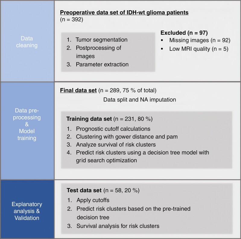
Methods: A total of 289 patients with glioblastoma who underwent preoperative multimodal MR imaging were included. Mean values of apparent diffusion coefficient normalized relative cerebral blood volume and relative cerebral blood flow were calculated for different tumor compartments and the entire tumor. Distinct imaging patterns were identified using partition around medoids (PAM) clustering on the training dataset, and their ability to predict overall survival was assessed. Additionally, tree-based machine-learning models were trained to ascertain the significance of features pertaining to cluster membership.
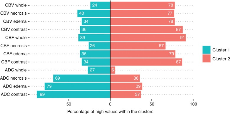
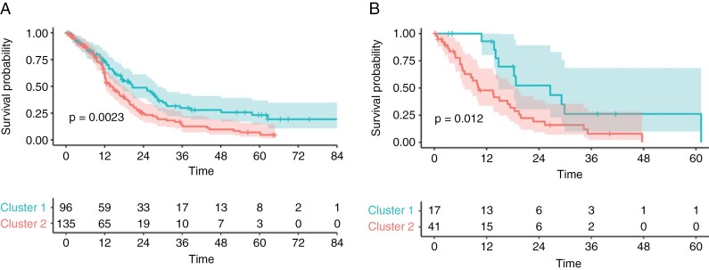
Results: Using the training dataset (231/289) we identified 2 stable imaging phenotypes through PAM clustering with significantly different overall survival (OS). Validation in an independent test set revealed a high-risk group with a median OS of 10.2 months and a low-risk group with a median OS of 26.6 months (P = 0.012). Patients in the low-risk cluster had high diffusion and low perfusion values throughout, while the high-risk cluster displayed the reverse pattern. Including cluster membership in all multivariate Cox regression analyses improved performance (P ≤ 0.004 each).
Conclusions: Our research demonstrates that data-driven clustering can identify clinically relevant, distinct imaging phenotypes, highlighting the potential role of diffusion, and perfusion MRI in predicting survival rates of glioblastoma patients.
Keywords: diffusion; glioblastoma; machine learning; perfusion; prognostic biomarker.
© The Author(s) 2023. Published by Oxford University Press on behalf of the Society for Neuro-Oncology. All rights reserved. For commercial re-use, please contact reprints@oup.com for reprints and translation rights for reprints. All other permissions can be obtained through our RightsLink service via the Permissions link on the article page on our site—for further information please contact journals.permissions@oup.com.
Conflict of interest statement
None declared.
Figures
Similar articles
-
Shape matters: unsupervised exploration of IDH-wildtype glioma imaging survival predictors.Eur Radiol. 2025 Mar;35(3):1351-1360. doi: 10.1007/s00330-024-11042-6. Epub 2024 Sep 9. Eur Radiol. 2025. PMID: 39251442 Free PMC article.
-
Ventricle contact is associated with lower survival and increased peritumoral perfusion in glioblastoma.J Neurosurg. 2018 Oct 19;131(3):717-723. doi: 10.3171/2018.5.JNS18340. J Neurosurg. 2018. PMID: 30485234
-
Apparent Diffusion Coefficient as a Predictive Biomarker for Survival in Patients with Treatment-Naive Glioblastoma Using Quantitative Multiparametric Magnetic Resonance Profiling.World Neurosurg. 2019 Feb;122:e812-e820. doi: 10.1016/j.wneu.2018.10.151. Epub 2018 Nov 1. World Neurosurg. 2019. PMID: 30391622
-
Clinical parameters outweigh diffusion- and perfusion-derived MRI parameters in predicting survival in newly diagnosed glioblastoma.Neuro Oncol. 2016 Dec;18(12):1673-1679. doi: 10.1093/neuonc/now122. Epub 2016 Jun 13. Neuro Oncol. 2016. PMID: 27298312 Free PMC article.
-
Multi-parametric and multi-regional histogram analysis of MRI: modality integration reveals imaging phenotypes of glioblastoma.Eur Radiol. 2019 Sep;29(9):4718-4729. doi: 10.1007/s00330-018-5984-z. Epub 2019 Feb 1. Eur Radiol. 2019. PMID: 30707277 Free PMC article.
Cited by
-
Shape matters: unsupervised exploration of IDH-wildtype glioma imaging survival predictors.Eur Radiol. 2025 Mar;35(3):1351-1360. doi: 10.1007/s00330-024-11042-6. Epub 2024 Sep 9. Eur Radiol. 2025. PMID: 39251442 Free PMC article.
-
Decoding Glioblastoma Heterogeneity: Neuroimaging Meets Machine Learning.Neurosurgery. 2025 Jun 1;96(6):1181-1192. doi: 10.1227/neu.0000000000003260. Epub 2024 Nov 21. Neurosurgery. 2025. PMID: 39570018 Review.
References
-
- Anzalone N, Castellano A, Cadioli M, et al.. Brain gliomas: multicenter standardized assessment of dynamic contrast-enhanced and dynamic susceptibility contrast MR Images. Radiology. 2018;287(3):933–943. - PubMed
Publication types
MeSH terms
Grants and funding
LinkOut - more resources
Full Text Sources
Medical
Miscellaneous
