

Diagnosis of Primary Ciliary Dyskinesia via Whole Exome Sequencing and Histologic Findings
- PMID: 38154480
- PMCID: PMC10774650
- DOI: 10.3349/ymj.2023.0238
Diagnosis of Primary Ciliary Dyskinesia via Whole Exome Sequencing and Histologic Findings
Abstract
Purpose: To assess the diagnostic potential of whole-exome sequencing (WES) and elucidate the clinical and genetic characteristics of primary ciliary dyskinesia (PCD) in the Korean population.
Materials and methods: Forty-seven patients clinically suspected of having PCD were enrolled at a tertiary medical center. WES was performed in all patients, and seven patients received biopsy of cilia and transmission electron microscopy (TEM).
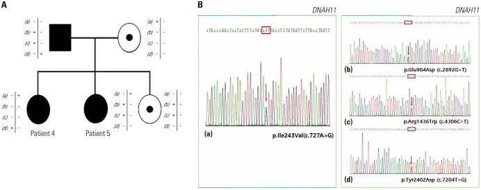
Results: Overall, PCD was diagnosed in 10 (21.3%) patients: eight by WES (8/47, 17%), four by TEM. Among patients diagnosed as PCD based on TEM results, two patients showed consistent results with WES and TEM of PCD (2/4, 50%). In addition, five patients, who were not included in the final PCD diagnosis group, had variants of unknown significance in PCD-related genes (5/47, 10.6%). The most frequent pathogenic (P)/likely pathogenic (LP) variants were detected in DNAH11 (n=4, 21.1%), DRC1 (n=4, 21.1%), and DNAH5 (n=4, 21.1%). Among the detected 17 P/LP variants in PCD-related genes in this study, 8 (47.1%) were identified as novel variants. Regarding the genotype-phenotype correlation in this study, the authors experienced severe PCD cases caused by the LP/P variants in MCIDAS, DRC1, and CCDC39.
Conclusion: Through this study, we were able to confirm the value of WES as one of the diagnostic tools for PCD, which increases with TEM, rather than single gene tests. These results will prove useful to hospitals with limited access to PCD diagnostic testing but with relatively efficient in-house or outsourced access to genetic testing at a pre-symptomatic or early disease stage.
Keywords: Primary ciliary dyskinesia; copy number variants analysis; genetic testing; transmission electron microscopy; whole exome sequencing.
© Copyright: Yonsei University College of Medicine 2024.
Conflict of interest statement
The authors have no potential conflicts of interest to disclose.
Figures
Similar articles
-
Genetic Analysis of Korean Adult Patients with Nontuberculous Mycobacteria Suspected of Primary Ciliary Dyskinesia Using Whole Exome Sequencing.Yonsei Med J. 2021 Mar;62(3):224-230. doi: 10.3349/ymj.2021.62.3.224. Yonsei Med J. 2021. PMID: 33635012 Free PMC article.
-
Genetics of 67 patients of suspected primary ciliary dyskinesia from India.Clin Genet. 2024 Nov;106(5):650-658. doi: 10.1111/cge.14590. Epub 2024 Jul 14. Clin Genet. 2024. PMID: 39004944
-
Ciliary Ultrastructure Assessed by Transmission Electron Microscopy in Adults with Bronchiectasis and Suspected Primary Ciliary Dyskinesia but Inconclusive Genotype.Cells. 2023 Nov 18;12(22):2651. doi: 10.3390/cells12222651. Cells. 2023. PMID: 37998386 Free PMC article.
-
Value of transmission electron microscopy for primary ciliary dyskinesia diagnosis in the era of molecular medicine: Genetic defects with normal and non-diagnostic ciliary ultrastructure.Ultrastruct Pathol. 2017 Nov-Dec;41(6):373-385. doi: 10.1080/01913123.2017.1362088. Epub 2017 Sep 15. Ultrastruct Pathol. 2017. PMID: 28915070 Free PMC article. Review.
-
A case of primary ciliary dyskinesis with DRC1 deletion and literature review: Additional evidence on the founder effect.Pediatr Int. 2024 Jan-Dec;66(1):e15808. doi: 10.1111/ped.15808. Pediatr Int. 2024. PMID: 39349394 Review.
References
-
- Lucas JS, Davis SD, Omran H, Shoemark A. Primary ciliary dyskinesia in the genomics age. Lancet Respir Med. 2020;8:202–216. - PubMed
-
- Kuehni CE, Frischer T, Strippoli MP, Maurer E, Bush A, Nielsen KG, et al. Factors influencing age at diagnosis of primary ciliary dyskinesia in European children. Eur Respir J. 2010;36:1248–1258. - PubMed
MeSH terms
LinkOut - more resources
Full Text Sources
Medical
Research Materials
