. 2023 Dec 27;13(1):23025.
doi: 10.1038/s41598-023-50365-0.

Identification of CD38, CD97, and CD278 on the HIV surface using a novel flow virometry screening assay

Affiliations

Identification of CD38, CD97, and CD278 on the HIV surface using a novel flow virometry screening assay

Jonathan Burnie et al. Sci Rep. .

Abstract

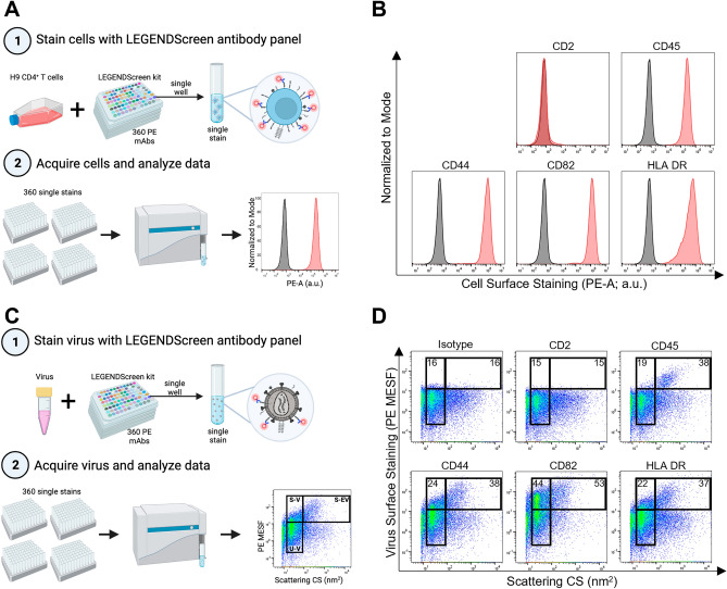
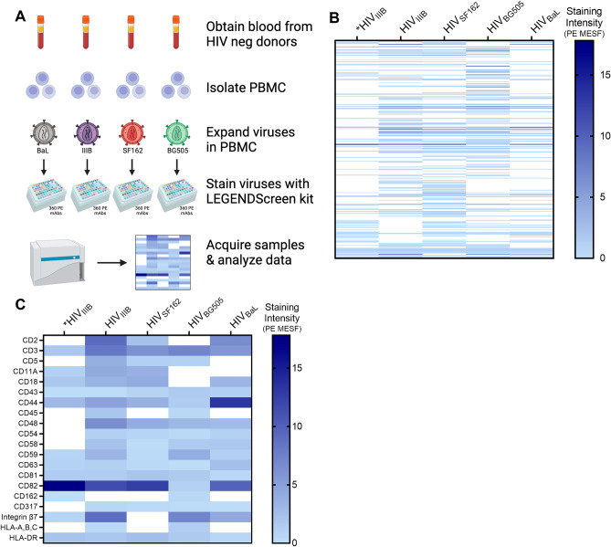
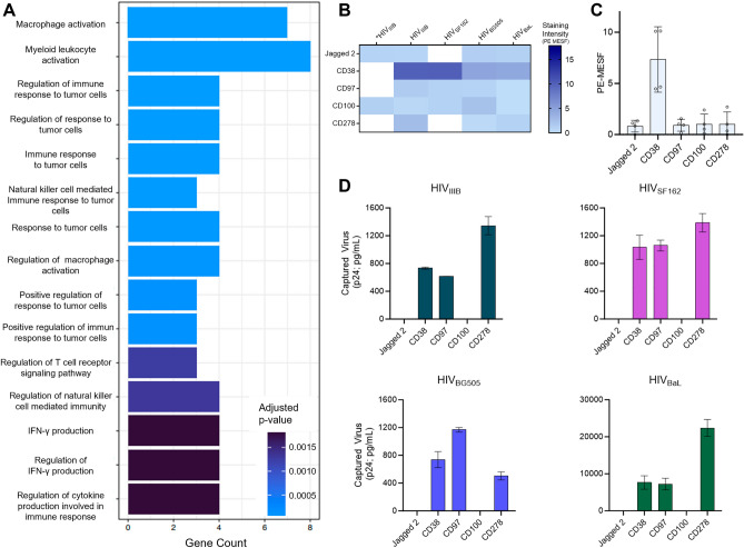
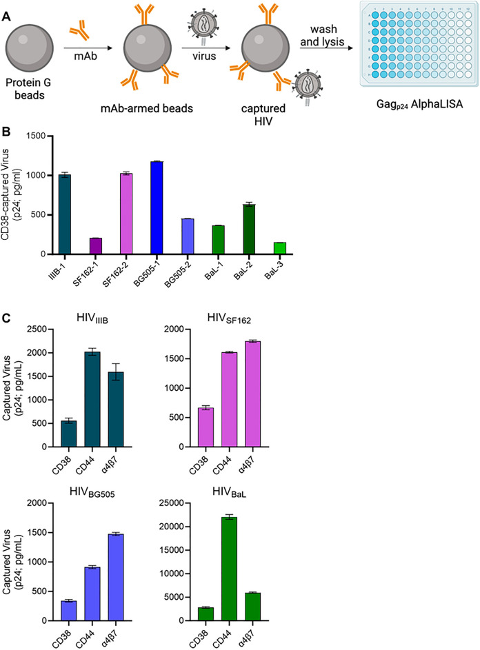
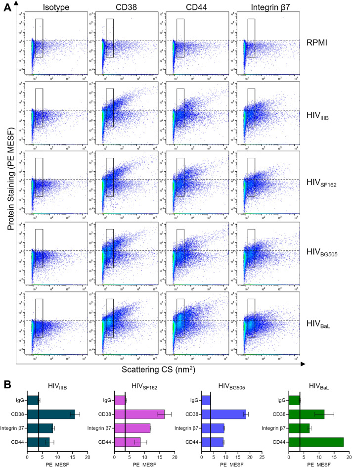
While numerous cellular proteins in the HIV envelope are known to alter virus infection, methodology to rapidly phenotype the virion surface in a high throughput, single virion manner is lacking. Thus, many human proteins may exist on the virion surface that remain undescribed. Herein, we developed a novel flow virometry screening assay to discover new proteins on the surface of HIV particles. By screening a CD4+ T cell line and its progeny virions, along with four HIV isolates produced in primary cells, we discovered 59 new candidate proteins in the HIV envelope that were consistently detected across diverse HIV isolates. Among these discoveries, CD38, CD97, and CD278 were consistently present at high levels on virions when using orthogonal techniques to corroborate flow virometry results. This study yields new discoveries about virus biology and demonstrates the utility and feasibility of a novel flow virometry assay to phenotype individual virions.


Conflict of interest statement

The authors declare no competing interests.

Figures

Figure 1
Figure 1
Adapting the LEGENDScreen kit for use on HIVIIIB and its producer cells. (A) Schematic depicting the experimental workflow used for cell staining. Step 1 shows infected H9 cells stained with 360 unique PE-labelled antibodies provided in the LEGENDScreen kit, which consists of four 96-well plates containing a single PE-labelled mAb clone in each well (different clones depicted as different colors). Each well was harvested into a single tube, as a single stain to be acquired in step 2. Step 2 shows stained cells acquired on the CytoFLEX nanoscale cytometer by flow cytometry methods. Data are displayed as fluorescence histograms with isotype staining shown in black and specific protein staining shown in red. (B) A representative subset of cell surface staining from the LEGENDScreen. (C) Schematic depicting the experimental workflow used for virus staining. Step 1 shows virus (HIVIIIB) produced in H9 cells that was stained with the LEGENDScreen antibodies and then transferred to tubes and diluted in PBS before proceeding to step 2 for acquisition. Data are displayed in pseudocolour dot plots to allow for the visualization of virus and vesicle populations. The lower gate denotes unstained virus (U-V). The left and right upper gates display stained virus (S-V) and stained extracellular vesicles (S-EV), respectively. (D) A representative subset of virus surface staining from the LEGENDScreen, with mean PE MESF values depicted in the gates, as shown. Data from one screen performed on the H9 cells and their respective viruses are shown.
Figure 2
Figure 2
Screening viruses from primary cells using a novel flow virometry assay. (A) Schematic depicting the experimental workflow used for staining PBMC viruses. Blood samples from four uninfected donors were used to isolate PBMC. Four different HIV isolates (BaL, IIIB, SF162, BG505) were propagated in PBMC. Each viral stock was then stained individually with the full panel of LEGENDScreen antibodies before being diluted with PBS, and then acquired on the cytometer. Data are displayed in a heatmap with darker shades representing higher levels of staining. (B) Heat map displaying the mean fluorescence (PE MESF) generated from each of the stained PBMC virus isolates that were tested (columns 2–5). Each row represents a different antibody stain from the LEGENDScreen panel, and each column represents a different viral isolate. Staining intensities for proteins of interest were subtracted from the mean staining values generated from staining with matched isotype controls. Samples with staining below the isotype control are shown in white. *Staining intensities from HIVIIIB produced in the H9 T cell line are shown in column one, whereas columns 2–5 show viral isolates propagated in PBMC. (C) A subset of antigens from (B) that were previously established to be present on HIV particles are labelled in (C), shown here as positive controls for assay specificity.
Figure 3
Figure 3
Characterizing novel virion-incorporated candidate proteins identified from the flow virometry screen. (A) Purported biological processes of the novel candidates from the LEGENDScreen as described through gene ontology analysis. Novel candidates were defined as proteins that have not been described in the literature on HIV, and those that stained above isotype on at least three out of four PBMC viruses tested. (B) A subset of novel candidate proteins identified through the LEGENDScreen. Each row represents a different antibody stain from the LEGENDScreen panel, and each column represents a different viral isolate. *Staining intensities from HIVIIIB produced in the H9 T cell line are shown in column one. Staining intensities for proteins of interest were subtracted from the mean staining values generated from staining with matched isotype controls. Samples with staining below the isotype control are shown in white. (C) Graphical representation of the PE MESF values observed in B for the selected novel proteins. Each dot represents a different viral isolate that was tested in the LEGENDScreen. Bars represent the mean PE MESF value ± standard deviation. MESF values with fluorescent intensity below the isotype controls were plotted at zero. (D) Virion capture assays were performed to corroborate the presence of the selected novel protein candidates on the viruses. Normalized inputs of the same virus stocks tested in the LEGENDScreen assays (HIVBaL, HIVIIIB, HIVBG505, HIVSF162) were incubated with plate-bound mAbs against Jagged 2, CD38, CD97, CD100, and CD278/ICOS. Captured virions were lysed and HIV Gag (p24) was quantified in the lysates (via AlphaLISA), as an indicator of the amount of virus capture. Values are representative of two independent experiments and are reported as the mean ± SD of duplicate AlphaLISA measurements. CD38, CD97, and CD278/ICOS were significantly higher than isotype capture (paired ANOVA; p = 0.001) across all four virus isolates. Virus capture values that were below the isotype control are shown as zero.
Figure 4
Figure 4
Bead-based capture of HIV virus stocks using an anti-CD38 antibody. (A) A schematic representation of the bead-based virion capture assay. Protein G coated magnetic beads are armed with the antibody (mAb) against the protein to be detected on the viral surface. The virus is incubated with the antibody-armed beads for 2 h at room temperature to allow virus capture, followed by washing and lysis of bead-bound (i.e., captured) virus. The amount of virus captured via each mAb is quantified by Gag p24 AlphaLISA performed on viral lysates. (B) Virion capture assays were performed on equal volumes of eight independent virus stocks of HIV isolates (BaL, IIIB, BG505, SF162) grown in different PBMC donors. The numbering of isolates indicates a distinct viral stock produced in a different PBMC donor for the same viral isolate (i.e., BaL-1, BaL-2, and BaL-3 are the same BaL isolate grown in 3 different PBMC donors). Bead-associated virus was lysed and HIV-1 p24 Gag was quantified using p24 AlphaLISA as an indicator of the amount of virus capture. Background levels of capture achieved with an isotype-matched control antibody were subtracted from all data shown. Levels of CD38 capture displayed are significantly higher than isotype capture (paired t-test; p = 0.003). (C) Virion capture assays were performed with immunomagnetic beads armed with antibodies against CD38, CD44 or integrin β7 with each virus isolate (BaL, IIIB, BG505, SF162). In parallel, we assessed the levels of background capture as measured with an isotype control, and these control values were subtracted from the capture data before graphing. Levels of CD38 capture displayed are significantly higher than isotype capture (paired ANOVA; p = 0.004). The mean ± SD are generated from independent virus stocks tested in duplicate.
Figure 5
Figure 5
Quantitative flow virometry staining of CD38 on the surface of HIV. (A) Four different HIV isolates (IIIB, BG505, SF162, BaL) produced in PBMC were stained with PE-conjugated antibodies against CD38, CD44 and integrin β7. A stained cell culture medium control (RPMI) is shown to assess the contribution of antibodies alone (in absence of virus) to background fluorescence. Gates demarcate where the majority of viruses are anticipated to fall based on light scattering. Dotted lines indicate the level of background PE fluorescence set based on the isotype control. (B) Quantitative representation of mean fluorescent data (PE MESF) ± SD generated through the gates in two experimental replicates from flow virometry data as in (A). Solid black lines indicate levels of background capture as measured with an isotype control. Data shown are representative of five replicates for each virus stock.

Similar articles

Cited by

References

    1. Tremblay MJ, Fortin J-F, Cantin R. The acquisition of host-encoded proteins by nascent HIV-1. Immunol. Today. 1998;19:346–351. doi: 10.1016/S0167-5699(98)01286-9. - DOI - PubMed
    1. Fortin J-F, Cantin R, Lamontagne G, Tremblay M. Host-derived ICAM-1 glycoproteins incorporated on human immunodeficiency virus type 1 are biologically active and enhance viral infectivity. J. Virol. 1997;71:3588–3596. doi: 10.1128/jvi.71.5.3588-3596.1997. - DOI - PMC - PubMed
    1. Saifuddin M. Role of virion-associated glycosylphosphatidylinositol-linked proteins CD55 and CD59 in complement resistance of cell line-derived and primary isolates of HIV-1. J. Exp. Med. 1995;182:501–509. doi: 10.1084/jem.182.2.501. - DOI - PMC - PubMed
    1. Bounou S, Leclerc JE, Tremblay MJ. Presence of host ICAM-1 in laboratory and clinical strains of human immunodeficiency virus type 1 increases virus infectivity and CD4+-T-cell depletion in human lymphoid tissue, a major site of replication in vivo. J. Virol. 2002;76:1004–1014. doi: 10.1128/JVI.76.3.1004-1014.2002. - DOI - PMC - PubMed
    1. Liao Z, Roos JW, Hildreth JEK. Increased infectivity of HIV type 1 particles bound to cell surface and solid-phase ICAM-1 and VCAM-1 through acquired adhesion molecules LFA-1 and VLA-4. AIDS Res. Hum. Retroviruses. 2000;16:355–366. doi: 10.1089/088922200309232. - DOI - PubMed