. 2024 May;244(5):803-814.
doi: 10.1111/joa.14000. Epub 2023 Dec 28.

Localisation of the centre of the highest region of muscle spindle abundance of anterior forearm muscles

Affiliations

Localisation of the centre of the highest region of muscle spindle abundance of anterior forearm muscles

Jiayu Zhou et al. J Anat. 2024 May.

Abstract

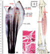
The centre of the highest region of muscle spindle abundance (CHRMSA) in the intramuscular nerve-dense region has been suggested as the optimal target location for injecting botulinum toxin A to block muscle spasms. The anterior forearm muscles have a high incidence of spasticity. However, the CHRMSA in the intramuscular nerve-dense region of the forearm anterior muscle group has not been defined. This study aimed to accurately define the body surface position and the depth of CHRMSA in an intramuscular nerve-dense region of the anterior forearm muscles. Twenty-four adult cadavers (57.7 ± 11.5 years) were included in this study. The curved line close to the skin connecting the medial and lateral epicondyles of the humerus was designated as the horizontal reference line (H line), and the line connecting the medial epicondyle of the humerus and the ulnar styloid was defined as the longitudinal reference line (L line). Modified Sihler's staining, haematoxylin-eosin staining and computed tomography scanning were employed to determine the projection points (P and P') of the CHRMSAs on the anterior and posterior surfaces of the forearm. The positions (PH and PL) of point P projected onto the H and L lines, and the depth of each CHRMSA, were determined using the Syngo system. The PH of the CHRMSA of the ulnar head of pronator teres, humeral head of pronator teres, flexor carpi radialis, palmaris longus, flexor carpi ulnaris, ulnar part of flexor digitorum superficialis, radial part of flexor digitorum superficialis, flexor pollicis longus, ulnar part of flexor digitorum profundus, radial portion of flexor digitorum profundus and pronator quadratus muscles were located at 42.48%, 45.52%, 41.20%, 19.70%, 7.77%, 25.65%, 47.42%, 53.47%, 12.28%, 38.41% and 51.68% of the H line, respectively; the PL were located at 18.38%, 12.54%, 28.83%, 13.43%, 17.65%, 32.76%, 57.32%, 64.12%, 20.05%, 45.94% and 88.71% of the L line, respectively; the puncture depths were located at 21.92%, 27.25%, 23.76%, 18.04%, 15.49%, 31.36%, 26.59%, 41.28%, 38.72%, 45.14% and 53.58% of the PP' line, respectively. The percentage values are the means of individual values. We recommend that the body surface puncture position and depth of the CHRMSA are the preferred locations for the intramuscular injection of botulinum toxin A to block anterior forearm muscle spasms.

Keywords: anterior forearm muscles; intramuscular nerve; muscle spindle abundance; spasm; target localization.


Conflict of interest statement

None.

Figures

FIGURE 1
FIGURE 1
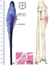
Sihler's staining of the right pronator teres muscle and representative muscle spindles in the highest region of muscle spindle abundance. (a) Sihler's staining showing the pattern of intramuscular nerve distribution (deep‐surface view). Scale bar, cm. The red frames represent the INDRs. The red dots represent the CHRMSAs. (b and c) Representative muscle spindles of the highest region of muscle spindle abundance in INDR1a and INDR1b; scale bar: 300 μm. (d) Schematic drawing of (a).
FIGURE 2
FIGURE 2
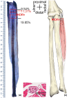
Sihler's staining of the right flexor carpi radialis muscle and representative muscle spindles in the highest region of muscle spindle abundance. (a) Sihler's staining showing the pattern of intramuscular nerve distribution (deep‐surface view). Scale bar, cm. The red box represents INDR2. The red dot represents the CHRMSA. (b) Representative muscle spindles of the highest region of muscle spindle abundance in INDR2. Scale, 300 μm. (c) Schematic drawing of (a).
FIGURE 3
FIGURE 3
Sihler's staining of the right palmaris longus muscle and representative muscle spindles in the highest region of muscle spindle abundance. (a) Sihler's staining showing the intramuscular nerve distribution pattern (deep‐surface view). Scale bar, cm. The red box represents INDR3. The red dot represents the CHRMSA. (b) Representative muscle spindle of the highest region of muscle spindle abundance in INDR3. Bar, 300 μm. (c) Schematic drawing of (a).
FIGURE 4
FIGURE 4
Sihler's staining of the right flexor carpi ulnaris muscle and representative muscle spindles in the highest region of muscle spindle abundance. (a) Sihler's staining showing the intramuscular nerve distribution pattern. Scale bar, cm. The red box represents INDR4. The red dot represents the CHRMSA. (b) Representative muscle spindle of the highest region of muscle spindle abundance in INDR4. scale bar, 300 μm. (c) Schematic drawing of (a).
FIGURE 5
FIGURE 5
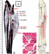
Sihler's staining of the right flexor digitorum superficialis muscle and representative muscle spindles in the highest region of muscle spindle abundance. (a) Sihler's staining showing the intramuscular nerve distribution pattern (deep‐surface view). Scale bar, cm. The red boxes represent the INDRs. The red dots represent the CHRMSAs. (b and c) Representative muscle spindles in the highest region of muscle spindle abundance in INDR5a and INDR5b, respectively. Bar, 300 μm. (d) Schematic drawing of (a).
FIGURE 6
FIGURE 6
Sihler's staining of the flexor pollicis longus muscle and representative muscle spindles in the highest region of muscle spindle abundance. (a) Sihler's staining showing the intramuscular nerve distribution pattern. Scale bar, cm. The red box represents INDR6. The red dot represents the CHRMSA. (b) Representative muscle spindles in the highest region of muscle spindle abundance. Bar, 300 μm. (c) Schematic drawing of (a).
FIGURE 7
FIGURE 7
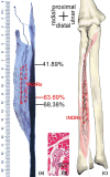
Sihler's staining of the right flexor digitorum profundus muscle and representative muscle spindles in the highest regions of muscle spindle abundance. (a) Sihler's staining showing the intramuscular nerve distribution pattern. Scale bar, cm. The red boxes represent the INDRs. The red dots represent the CHRMSAs. (b and c) Representative muscle spindles in the highest region of muscle spindle abundance for INDR7a and INDR7b, respectively. Bar, 300 μm. (d) Schematic drawing of (a).
FIGURE 8
FIGURE 8
Sihler's staining of the pronator quadratus muscle and representative muscle spindles in the highest region of muscle spindle abundance. (a) Sihler's staining showing the intramuscular nerve distribution pattern (deep‐surface view). Scale bar, cm. The red box represents INDR8. The red dot represents the CHRMSA. (b) Representative muscle spindle of the highest region of muscle spindle abundance in INDR8. Bar, 300 μm. (c) Schematic drawing of (a).
FIGURE 9
FIGURE 9
Spiral CT localisation image of the CHRMSA of the left flexor pollicis longus muscle. (a) Spiral CT localisation image showing the position of each CHRMSA of the anterior forearm muscles on the body surface and the designed reference line. (a1) anterior forearm, (a2) posterior forearm. a—medial epicondyle of the humerus; b—lateral epicondyle of the humerus; c—ulnar styloid process. P6—the body surface projection point of the CHRMSA of the flexor pollicis longus muscle. P6H—the intersection of the vertical line through P6 and the H line. P6L—the intersection of the horizontal line through P6 and the L line. a‐P6H=H6’, a‐P6L = L6’. (b) Measurement of the length of the L and L6’ lines on the coronal section. (c) Measurement of the lengths of the H and H6’ lines on the cross section. (d) Measurement of the depth of CHRMSA6 on the cross section.

References

    1. Abo, M. , Shigematsu, T. , Hara, H. , Matsuda, Y. , Nimura, A. , Yamashita, Y. et al. (2020) Efficacy and safety of onabotulinumtoxina 400 units in patients with post‐stroke upper limb spasticity: final report of a randomized, double‐blind, placebo‐controlled trial with an open‐label extension phase. Toxins (Basel), 12(2), 127. - PMC - PubMed
    1. Akkaya, T. , Unlu, E. , Alptekin, A. , Gumus, H.I. , Umay, E. & Cakci, A. (2010) Neurolytic phenol blockade of the obturator nerve for severe adductor spasticity. Acta Anaesthesiologica Scandinavica, 54(1), 79–85. - PubMed
    1. Andresen, R. , Radmer, S. , Nickel, J. , Fischer, G. & Brinckmann, W. (2009) Ambulante CT‐gestützte thorakale Sympathikusblockade als zusätzliche Therapieoption bei komplexem regionalen Schmerzsyndrom Typ I nach Sportverletzungen [Ambulatory CT‐assisted thoracic sympathetic block as an additional approach to treatment of complex regional pain syndromes after sport injuries]. Sportverletzung Sportschaden, 23(1), 35–40. - PubMed
    1. Banks, R.W. (2006) An allometric analysis of the number of muscle spindles in mammalian skeletal muscles. Journal of Anatomy, 208(6), 753–768. - PMC - PubMed
    1. Borodic, G.E. , Ferrante, R. , Pearce, L.B. & Smith, K. (1994) Histologic assessment of dose‐related diffusion and muscle fiber response after therapeutic botulinum A toxin injections. Movement Disorders, 9(1), 31–39. - PubMed

Publication types

Substances