Randomized Controlled Trial
. 2024 Feb;11(2):263-277.
doi: 10.1002/acn3.51949. Epub 2023 Dec 28.

Infarct-related structural disconnection and delirium in surgical aortic valve replacement patients

Collaborators, Affiliations
Randomized Controlled Trial

Infarct-related structural disconnection and delirium in surgical aortic valve replacement patients

Jeffrey N Browndyke et al. Ann Clin Transl Neurol. 2024 Feb.

Abstract

Objective: Although acute brain infarcts are common after surgical aortic valve replacement (SAVR), they are often unassociated with clinical stroke symptoms. The relationship between clinically "silent" infarcts and in-hospital delirium remains uncertain; obscured, in part, by how infarcts have been traditionally summarized as global metrics, independent of location or structural consequence. We sought to determine if infarct location and related structural connectivity changes were associated with postoperative delirium after SAVR.

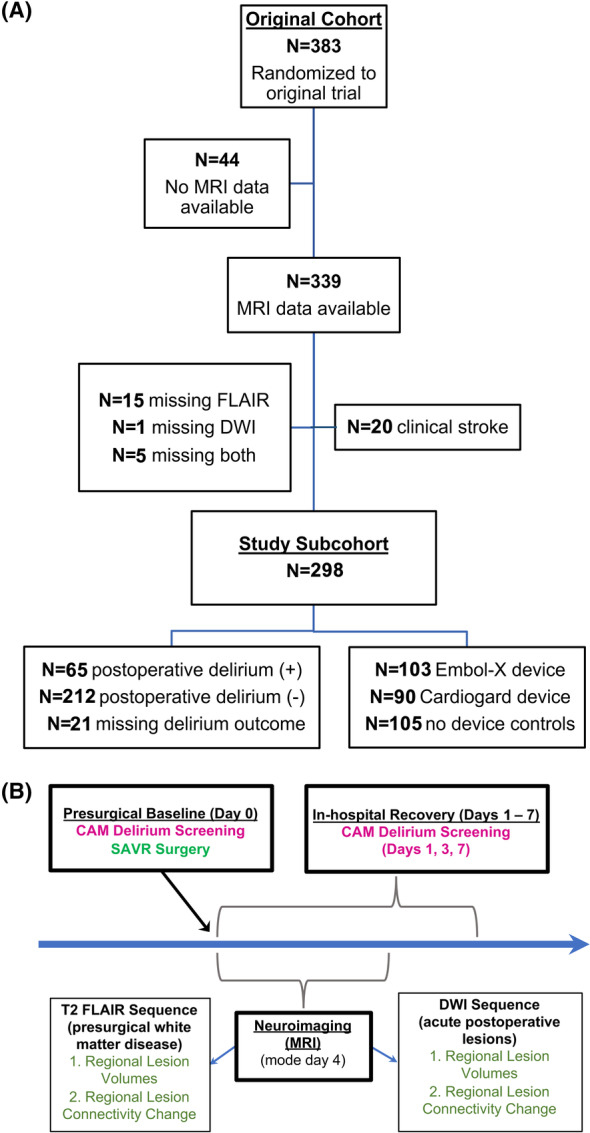
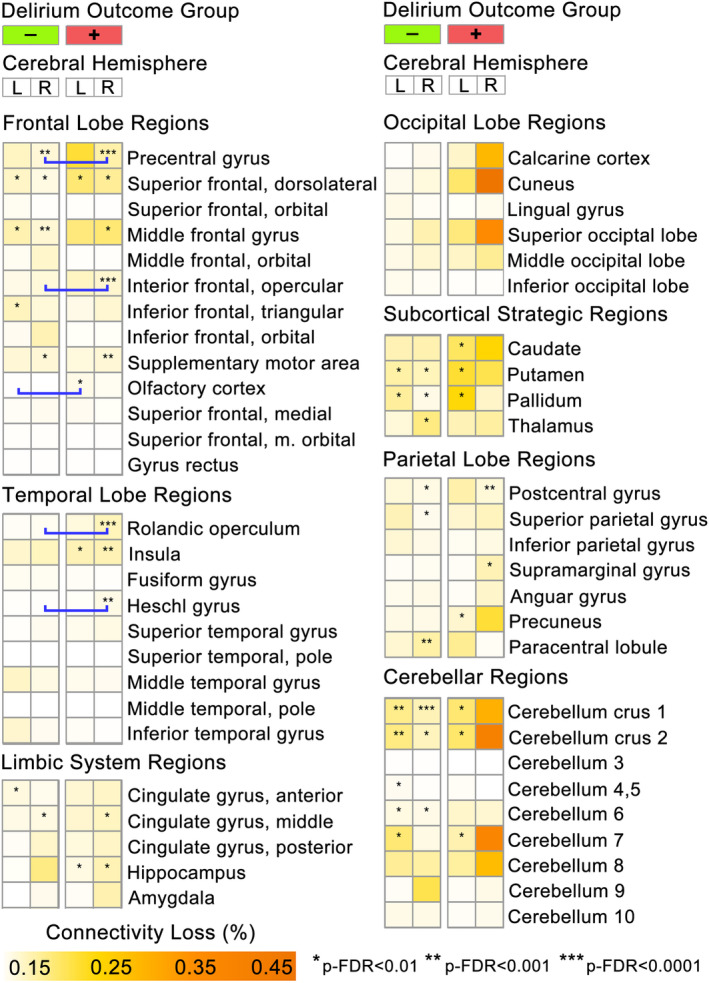
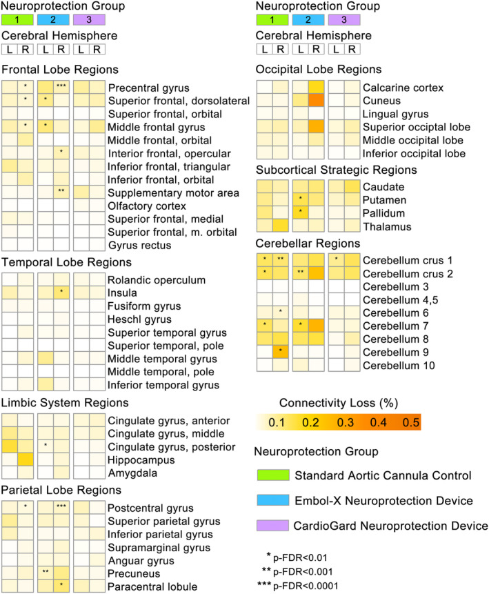
Methods: A secondary analysis of a randomized multicenter SAVR trial of embolic protection devices (NCT02389894) was conducted, excluding participants with clinical stroke or incomplete neuroimaging (N = 298; 39% female, 7% non-White, 74 ± 7 years). Delirium during in-hospital recovery was serially screened using the Confusion Assessment Method. Parcellation and tractography atlas-based neuroimaging methods were used to determine infarct locations and cortical connectivity effects. Mixed-effect, zero-inflated gaussian modeling analyses, accounting for brain region-specific infarct characteristics, were conducted to examine for differences within and between groups by delirium status and perioperative neuroprotection device strategy.

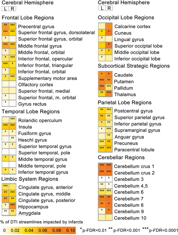
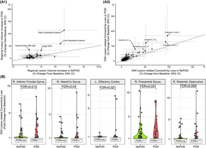
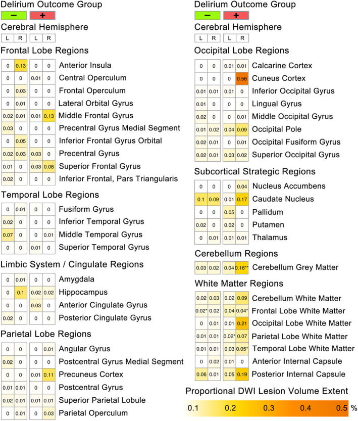
Results: 23.5% participants experienced postoperative delirium. Delirium was associated with significantly increased lesion volumes in the right cerebellum and temporal lobe white matter, while diffusion weighted imaging infarct-related structural disconnection (DWI-ISD) was observed in frontal and temporal lobe regions (p-FDR < 0.05). Fewer brain regions demonstrated DWI-ISD loss in the suction-based neuroprotection device group, relative to filtration-based device or standard aortic cannula.

Interpretation: Structural disconnection from acute infarcts was greater in patients who experienced postoperative delirium, suggesting that the impact from covert perioperative infarcts may not be as clinically "silent" as commonly assumed.


Conflict of interest statement

None of the authors have commercial relationships with the companies whose products were used in the study or may be affected by its outcomes.

Figures

Figure 1
Figure 1
Study CONSORT diagram and schematic of trial operations and outcome timing. (A) CONSORT diagram demonstrating the flow of patients from the original parent trial through to the current image‐based study sub‐cohort. (B) Illustrates the trial design, including timing of interventions and outcome measures.
Figure 2
Figure 2
Regional perioperative acute ischemic DWI infarct‐mediated cortical disconnectivity in all groups. Heatmap of cortical disconnectivity associated with acute ischemic diffusion‐weighted imaging (DWI) lesions for each ROI, separated by cerebral hemisphere. Measured using change in connectivity (ChaCo) values, which reflect the % of afferent DTI streamlines impacted within each region‐of‐interest (ROI). Asterisks indicate significant change in connectivity from presurgical baseline; *p‐FDR < 0.01, **p‐FDR < 0.001, ***p‐FDR < 0.0001.
Figure 3
Figure 3
Comparing SAVR‐induced infarct volume and connectivity loss between patients with and without POD. Scatterplots show average percent change (95% CI) from presurgical baseline in lesion volume (A.1) or DWI lesion‐related connectivity loss (A.2) for each ROI in patients with POD (y‐axis) versus without POD (x‐axis). Labels indicate ROIs with POD‐change minus NoPOD‐change >5% (A.1; right cerebellum and temporal lobe ROIs significantly different between POD groups, p‐FDR < 0.05). The dashed line represents the origin line. Violin plots (B) show DWI lesion‐related connectivity loss change from presurgical baseline within delirium groups for ROIs with significant group‐wise differences (p‐FDR < 0.05). Within group change significance denoted as p‐FDR: *<0.01, **<0.001, ***<0.0001.
Figure 4
Figure 4
Regional perioperative diffusion‐weighted imaging (DWI) lesion extent by cerebral hemisphere and delirium outcome group. Heatmap of regional perioperative diffusion‐weighted imaging (DWI) lesion volumes corrected for individual ROI volumes (i.e., DWI lesion volume/ROI volume = regional lesion extent), separated by cerebral hemisphere and delirium outcome group. ROIs with lesion extent >0.01% are shown. Asterisks indicate significant change in regional lesion extent from presurgical baseline; *p‐FDR < 0.01, **p‐FDR < 0.001, ***p‐FDR < 0.0001.
Figure 5
Figure 5
Significant SAVR‐associated DWI infarct changes in structural connectivity are more frequently observed in patients who experienced postoperative delirium. Heatmap shows the mean perioperative DWI lesion‐mediated connectivity loss (DWI‐ISD) for each ROI, separated by hemisphere and delirium outcome. Stars indicate ROIs with significant within‐group fold‐change in SAVR‐induced connectivity loss from presurgical baseline (p‐FDR corrected). Blue bracket bars indicate ROIs with significant differences (p‐FDR < 0.05) in DWI‐ISD between POD and no POD groups (see Fig. 3).
Figure 6
Figure 6
Reduced DWI infarct‐mediated structural connectivity loss with embolic capture neuroprotection device use during SAVR. Brain region heatmap shows the mean perioperative DWI lesion‐mediated connectivity loss for each ROI, separated by hemisphere and surgical device group. Stars indicate ROIs with significant within‐group fold‐change in connectivity loss from presurgical baseline (p‐FDR < 0.01).

References

    1. Messe SR, Acker MA, Kasner SE, et al. Stroke after aortic valve surgery: results from a prospective cohort. Circulation. 2014;129(22):2253‐2261. doi:10.1161/CIRCULATIONAHA.113.005084 - DOI - PMC - PubMed
    1. Samim M, Hendrikse J, van der Worp HB, et al. Silent ischemic brain lesions after transcatheter aortic valve replacement: lesion distribution and predictors. Clin Res Cardiol. 2015;104(5):430‐438. doi:10.1007/s00392-014-0798-8 - DOI - PubMed
    1. Stolz E, Gerriets T, Kluge A, Klovekorn WP, Kaps M, Bachmann G. Diffusion‐weighted magnetic resonance imaging and neurobiochemical markers after aortic valve replacement: implications for future neuroprotective trials? Stroke. 2004;35(4):888‐892. doi:10.1161/01.STR.0000120306.82787.5A - DOI - PubMed
    1. Evered L, Silbert B, Knopman DS, et al. Recommendations for the nomenclature of cognitive change associated with anaesthesia and surgery‐2018. J Alzheimers Dis. 2018;66(1):1‐10. doi:10.3233/JAD-189004 - DOI - PubMed
    1. Shadvar K, Baastani F, Mahmoodpoor A, Bilehjani E. Evaluation of the prevalence and risk factors of delirium in cardiac surgery ICU. J Cardiovasc Thorac Res. 2013;5(4):157‐161. doi:10.5681/jcvtr.2013.034 - DOI - PMC - PubMed

Publication types