. 2024 Mar;23(3):e14075.
doi: 10.1111/acel.14075. Epub 2023 Dec 28.

Phosphodiesterase inhibitor ameliorates senescent changes of renal interstitial pericytes in aging kidney

Affiliations

Phosphodiesterase inhibitor ameliorates senescent changes of renal interstitial pericytes in aging kidney

Hyung Duk Kim et al. Aging Cell. 2024 Mar.

Abstract

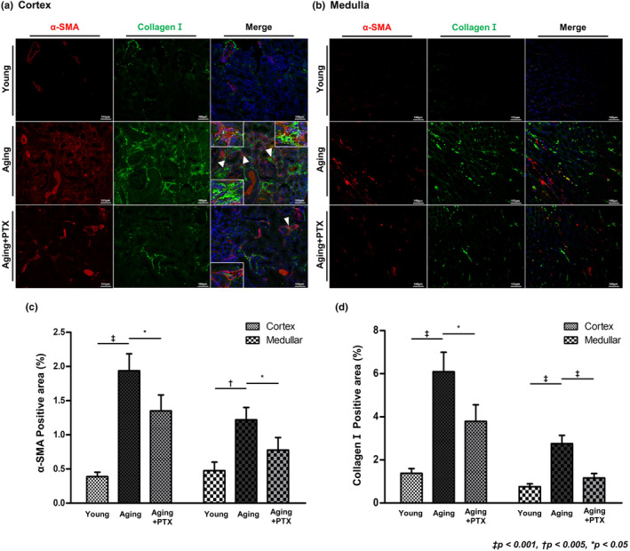
Pericytes are mesenchymal cells that surround endothelial cells, playing a crucial role in angiogenesis and vessel maturation. Additionally, they are associated with interstitial fibrosis as a major contributor to renal myofibroblasts. In this study, we aim to investigate whether the phosphodiesterase inhibitor, pentoxifylline (PTX), can ameliorate aging-related functional and histological deterioration in the kidney. We subjected aging C57BL/6 mice, dividing into young, aging, and PTX-treated aging groups. Renal function, albuminuria, and histological changes were assessed. Interstitial pericytes were assessed by immunohistochemistry analysis. We examined changes in pericytes in elderly patients using human kidney tissue obtained from healthy kidney donors for kidney transplantation. In vitro experiments with human pericytes and endothelial cells were performed. Aging mice exhibited declined renal function, increased albuminuria, and aging-related histological changes including mesangial expansion and tubulointerstitial fibrosis. Notably, number of pericytes declined in aging kidneys, and myofibroblasts increased. PTX treatment ameliorated albuminuria, histological alterations, and microvascular rarefaction, as well as modulated angiopoietin expression. In vitro experiments showed PTX reduced cellular senescence and inflammation. Human kidney analysis confirmed similar pericyte changes in aging kidneys. The phosphodiesterase inhibitor, PTX preserved microvascular density and improved renal interstitial fibrosis and inflammation in aging mice kidneys. These protective effects were suggested to be associated with the amelioration of pericytes reduction and the transition to myofibroblasts. Additionally, the upregulation of angiopoietin-1 expression may exert potential impacts. To the best of our knowledge, this is the first report on the changes in renal interstitial pericytes in aging human kidneys.

Keywords: aging; microvascular rarefaction; pentoxifylline; pericytes; phosphodiesterase inhibitors.


Conflict of interest statement

The authors declare that the research was conducted in the absence of any commercial or financial relationships that could be construed as a potential conflict of interest.

Figures

FIGURE 1
FIGURE 1
Study protocol and effects of pentoxifylline (PTX) on renal function and aging‐related histological renal injuries. (a) Schematic representation of the study protocol. Baseline samples including serum and urine were obtained at the 18th month. PTX was administered for 6 months at a dosage of 20 mg/kg/day in PTX group. The experimental animals were sacrificed at the 24th month, and serum, urine, and kidney tissue samples were obtained for analysis. Compared to the aging group, PTX group showed (b) lower albuminuria. PTX group showed (c) lower serum creatinine and (d) better creatinine clearance, though not statistically significant. (e) Representative photomicrographs of the periodic acid‐Schiff‐(PAS)‐stained kidney showed less expansion of the mesangial area in PTX group (original magnification, 400×). (f) Representative sections of the Masson's trichrome‐stained kidney showed significantly less tubulointerstitial fibrosis in PTX group (original magnification, 200×). Quantitative assessments of (g) the areas of extracellular matrix in the glomerulus and (h) the areas of tubulointerstitial fibrosis. (N ≥ 7 for all experiments. p < 0.001, p < 0.005, *p < 0.05).
FIGURE 2
FIGURE 2
Effects of pentoxifylline (PTX) on interstitial fibrosis and microvascular density. Interstitial fibrosis was assessed by immunofluorescence staining for collagen I (green) and microvascular density was measured by immunofluorescence staining for CD31 (red) in the (a) renal cortex and (b) medulla. (c) Quantitative analyses of CD31‐positive cells. (N ≥ 7 for all experiments. p < 0.005, *p < 0.05).
FIGURE 3
FIGURE 3
Effects of pentoxifylline (PTX) on interstitial fibrosis and expression of myofibroblasts. Interstitial fibrosis was assessed by immunofluorescence staining for collagen I (green) and myofibroblast was measured by immunofluorescence staining for α‐SMA (red) in (a) renal cortex and (b) medulla. (c, d) Quantitative analyses of the results. (N ≥ 7 for all experiments. p < 0.001, p < 0.005, *p < 0.05).
FIGURE 4
FIGURE 4
Effects of pentoxifylline (PTX) on interstitial pericytes. (a) Interstitial pericytes were identified by double staining of PDGFR‐β and NG2 (green and blue, respectively) and peritubular capillaries were identified by CD31 staining (red). (b) The number of pericytes was decreased in the aging group compared to that in the young group. Compared to the aging group, PTX group showed increased number of pericytes. (c) The number of pericytes surrounding peritubular capillaries was decreased in the aging group than in the young group. Compared to the aging group, PTX group showed increased number of pericytes surrounding peritubular capillaries. (N ≥ 7 for all experiments. p < 0.001, p < 0.005, *p < 0.05).
FIGURE 5
FIGURE 5
Effects of pentoxifylline (PTX) on the subset of pericytes differentiating into myofibroblasts. (a) Myofibroblasts were identified by α‐SMA staining. Co‐staining of α‐SMA and PDGFR‐β or α‐SMA and NG2 confirmed the presence of myofibroblasts derived from interstitial pericytes. (b–f) Cells with co‐staining of α‐SMA and PDGFR‐β/ α‐SMA and NG2/ α‐SMA, PDGFR‐β, and NG2 were increased in the aging group than in the young group, and decreased in the PTX group than in the aging group. (N ≥ 7 for all experiments. p < 0.001, p < 0.005, *p < 0.05).
FIGURE 6
FIGURE 6
Representative western blots of angiopoietins and inflammatory cytokines. (a) In western blot analysis, PTX group showed increased expression of angiopoietin‐1 and decreased expression of angiopoietin‐2, TNF‐α, IL‐6, and IL‐1β, compared to the aging group. (b–f) Quantitative analyses of the results. (N ≥ 7 for all experiments. p < 0.001, p < 0.005, *p < 0.05).
FIGURE 7
FIGURE 7
Doxorubicin‐induced cellular senescence and the expression of angiopoietins. Representative images of PTX treatment in (a) senescence‐induced pericytes and (b) HuVECs. (c) Pericytes and HuVECs treated with 200 nM doxorubicin showed increase in the number of β‐galactosidase‐positive cells, which was ameliorated with PTX treatment. (d) Representative western blots demonstrating angiopoietin‐1 and TNF‐α expression in senescence‐induced pericytes. In senescence‐induced pericytes, (e) angiopoietin‐1 expression was increased and (f) TNF‐α expression was decreased with PTX treatment. (g) Representative western blots demonstrating angiopoietin‐2 and TNF‐α expression in senescence‐induced HuVECs. (h, i) In senescence‐induced HuVECs, angiopoietin‐2 and TNF‐α expression was decreased with PTX treatment. (N ≥ 7 for all experiments. p < 0.001, p < 0.005, *p < 0.05).
FIGURE 8
FIGURE 8
Tubulointerstitial fibrosis and interstitial pericytes in human kidney. (a) Representative sections of the Masson's trichrome‐stained kidney showed significantly less tubulointerstitial fibrosis in the young group than in the aging group (original magnification, 200×). Interstitial pericytes were identified by double staining of PDGFR‐β and NG2 (green and blue, respectively) and peritubular capillaries were identified by CD31 staining (red). (b) Quantitative assessments of the areas of tubulointerstitial fibrosis. (c) The number of pericytes surrounding peritubular capillaries was decreased in the aging group than in the young group. (d) In elderly patients, tubulointerstitial fibrosis was increased and interstitial pericytes were decreased than in the young patients. (N = 12 for both groups. p < 0.001, *p < 0.05).

References

    1. Armulik, A. , Genové, G. , & Betsholtz, C. (2011). Pericytes: Developmental, physiological, and pathological perspectives, problems, and promises. Developmental Cell, 21(2), 193–215. 10.1016/j.devcel.2011.07.001 - DOI - PubMed
    1. Berthiaume, A. A. , Schmid, F. , Stamenkovic, S. , Coelho‐Santos, V. , Nielson, C. D. , Weber, B. , Majesky, M. W. , & Shih, A. Y. (2022). Pericyte remodeling is deficient in the aged brain and contributes to impaired capillary flow and structure. Nature Communications, 13(1), 5912. 10.1038/s41467-022-33464-w - DOI - PMC - PubMed
    1. Brindle, N. P. , Saharinen, P. , & Alitalo, K. (2006). Signaling and functions of angiopoietin‐1 in vascular protection. Circulation Research, 98(8), 1014–1023. 10.1161/01.Res.0000218275.54089.12 - DOI - PMC - PubMed
    1. Cai, J. , Kehoe, O. , Smith, G. M. , Hykin, P. , & Boulton, M. E. (2008). The angiopoietin/Tie‐2 system regulates pericyte survival and recruitment in diabetic retinopathy. Investigative Ophthalmology & Visual Science, 49(5), 2163–2171. 10.1167/iovs.07-1206 - DOI - PubMed
    1. Chang, F. C. , Chiang, W. C. , Tsai, M. H. , Chou, Y. H. , Pan, S. Y. , Chang, Y. T. , Yeh, P. Y. , Chen, Y. T. , Chiang, C. K. , Chen, Y. M. , Chu, T. S. , Wu, K. D. , & Lin, S. L. (2014). Angiopoietin‐2‐induced arterial stiffness in CKD. Journal of the American Society of Nephrology: JASN, 25(6), 1198–1209. 10.1681/asn.2013050542 - DOI - PMC - PubMed

Publication types

Substances