

Direct Observation of In-Focus Plasmonic Cargos via Breaking Angular Degeneracy in Differential Interference Contrast Microscopy
- PMID: 38155657
- PMCID: PMC10751767
- DOI: 10.1021/jacsau.3c00594
Direct Observation of In-Focus Plasmonic Cargos via Breaking Angular Degeneracy in Differential Interference Contrast Microscopy
Abstract
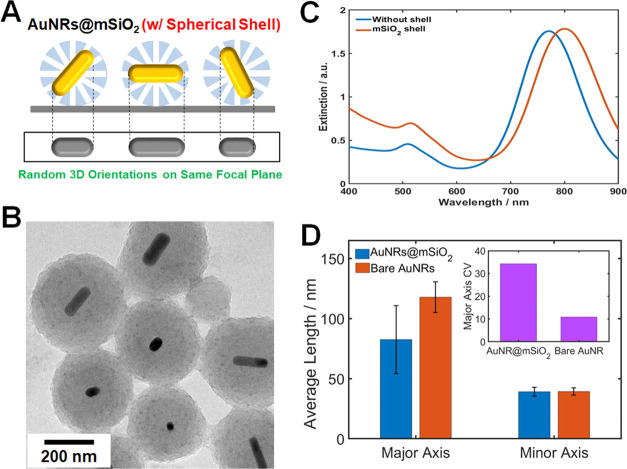
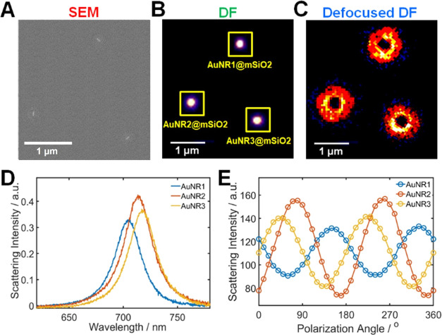
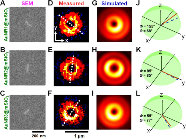
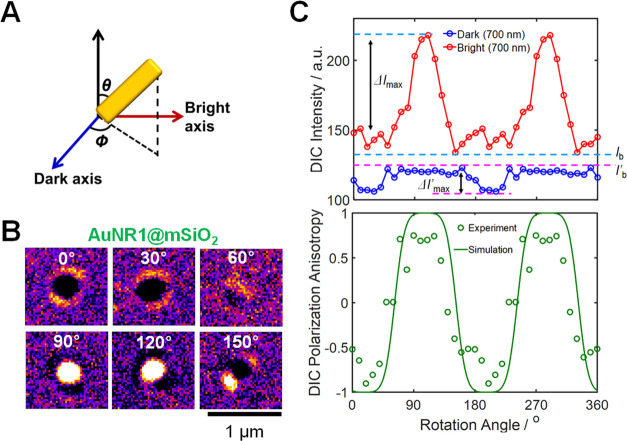
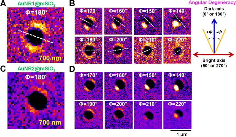
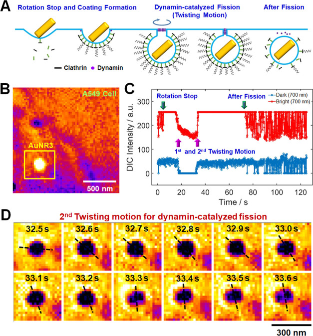
Breaking the angular degeneracy arising from the 2-fold optical symmetry of plasmonic anisotropic nanoprobes is critical in biological studies. In this study, we propose differential interference contrast (DIC) microscopy-based focused orientation and position imaging (dFOPI) to break the angular degeneracy of single gold nanorods (AuNRs). Single in-focus AuNRs (39 nm × 123 nm) within a spherical mesoporous silica shell were characterized with high throughput and produced distinct doughnut-shaped DIC image patterns featuring two lobes in the peripheral region, attributed to the scattering contribution of the AuNRs with large scattering cross sections. Interestingly, rotation of the lobes was observed in the focal plane for a large AuNR (>100 nm) tilted by more than ∼20° from the horizontal plane as the rotational stage was moved by 10° in a rotational study. From the rotation-dependent characteristic patterns, we directly visualized counterclockwise/clockwise rotations without the angular degeneracy at the localized surface plasmon resonance wavelength. Therefore, our dFOPI method can be applied for in vivo studies of important biological systems. To validate this claim, we tracked the three-dimensional rotational behavior of transferrin-modified in-focus AuNRs during clathrin-mediated endocytosis in real time without sacrificing the temporal and spatial resolution. In the invagination and scission stage, one or two directed twist motions of the AuNR cargos detached the AuNR-containing vesicles from the cell membrane. Furthermore, the dFOPI method directly visualized and revealed the right-handed twisting action along the dynamin helix in dynamin-catalyzed fission in live cells.
© 2023 The Authors. Published by American Chemical Society.
Conflict of interest statement
The authors declare no competing financial interest.
Figures
Similar articles
-
Focused orientation and position imaging (FOPI) of single anisotropic plasmonic nanoparticles by total internal reflection scattering microscopy.Nano Lett. 2012 Aug 8;12(8):4282-8. doi: 10.1021/nl301972t. Epub 2012 Jul 17. Nano Lett. 2012. PMID: 22793645
-
High-throughput in-focus differential interference contrast imaging of three-dimensional orientations of single gold nanorods coated with a mesoporous silica shell.RSC Adv. 2020 Aug 12;10(50):29868-29872. doi: 10.1039/d0ra04704j. eCollection 2020 Aug 10. RSC Adv. 2020. PMID: 35518257 Free PMC article.
-
Mesoporous silica shell-coated single gold nanorods as multifunctional orientation probes in dynamic biological environments.RSC Adv. 2021 Dec 1;11(61):38632-38637. doi: 10.1039/d1ra06572f. eCollection 2021 Nov 29. RSC Adv. 2021. PMID: 35493222 Free PMC article.
-
Gold Nanorods for LSPR Biosensing: Synthesis, Coating by Silica, and Bioanalytical Applications.Biosensors (Basel). 2020 Oct 17;10(10):146. doi: 10.3390/bios10100146. Biosensors (Basel). 2020. PMID: 33080925 Free PMC article. Review.
-
Individual Plasmonic Nanoprobes for Biosensing and Bioimaging: Recent Advances and Perspectives.Small. 2021 Feb;17(8):e2004287. doi: 10.1002/smll.202004287. Epub 2021 Feb 1. Small. 2021. PMID: 33522074 Review.
References
LinkOut - more resources
Full Text Sources