Review
. 2023 Dec 7;27(1):108683.
doi: 10.1016/j.isci.2023.108683. eCollection 2024 Jan 19.

Signaling pathways of liver regeneration: Biological mechanisms and implications

Affiliations
Review

Signaling pathways of liver regeneration: Biological mechanisms and implications

Chunyan Zhang et al. iScience. .

Abstract

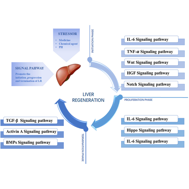
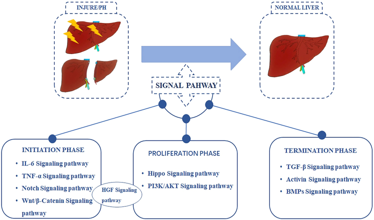
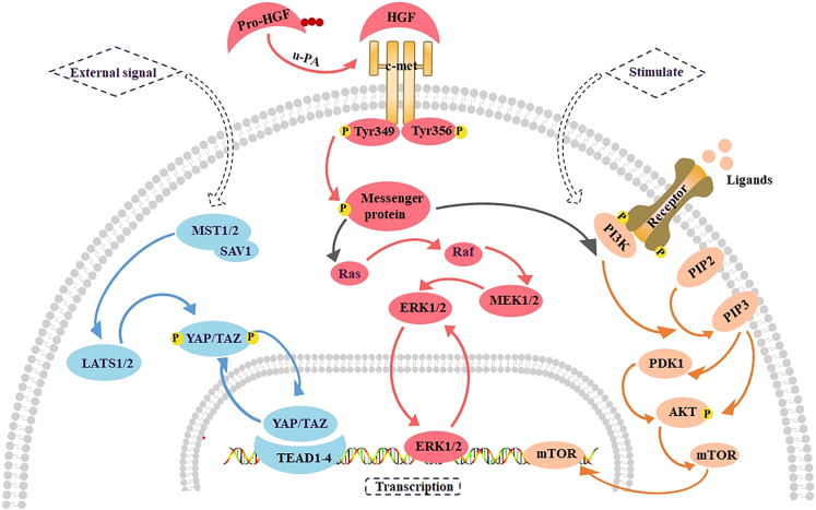
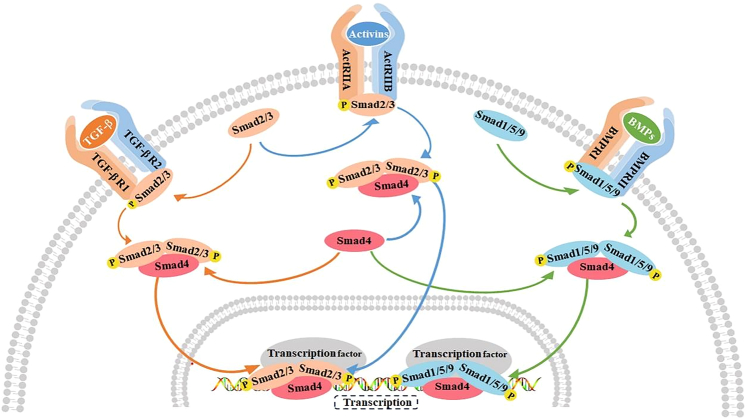
The liver possesses a unique regenerative ability to restore its original mass, in this regard, partial hepatectomy (PHx) and partial liver transplantation (PLTx) can be executed smoothly and safely, which has important implications for the treatment of liver disease. Liver regeneration (LR) can be the very complicated procedure that involves multiple cytokines and transcription factors that interact with each other to activate different signaling pathways. Activation of these pathways can drive the LR process, which can be divided into three stages, namely, the initiation, progression, and termination stages. Therefore, it is important to investigate the pathways involved in LR to elucidate the mechanism of LR. This study reviews the latest research on the key signaling pathways in the different stages of LR.

Keywords: Biological sciences; Health sciences.


Conflict of interest statement

The authors declare no competing interests.

Figures

None
Graphical abstract
Figure 1
Figure 1
The main activated signaling pathways in LR KCs and HSCs secrete TGFβ1 and activate TGF-β signaling pathway to act on SECs and hepatocytes. KCs and HSCs secrete HGF and activate HGF Signaling pathway to act on hepatocytes. KCs secrete TNF-α, IL-6 and activate TNF-α and IL-6 signaling pathway to act on hepatocytes. KCs and SECs secrete Wnt and activate Wnt signaling pathway to act on hepatocytes. HSCs and hepatocytes secrete pro-inflammatory cytokine activating activin A and activin signaling pathway that acts on HSCs and hepatocytes. HSCs, Hepatic stellate cells; KCs, Kupffer cells; SECs, Hepatic sinusoidal endothelial cells; HGF, Hepatocyte growth factor; TG.F-β, Transforming growth factor β; TNF-α, Tumor necrosis factor-α; IL-6, Interleukin-6. Dotted line arrow represents activation between cytokines.
Figure 2
Figure 2
The main signaling pathways at each phase of LR According to the initiation, progression and termination stages of LR, the relevant signaling pathways in the process of LR can be divided into three stages. Some signaling pathways can be involved in different stages of LR. PHx, Partial hepatectomy; PI3K, Phosphoinositide 3-kinase; BMPs, bone morphogenetic proteins.
Figure 3
Figure 3
The main signaling pathways in the initiation phase of LR Wnt, HGF, IL-6, TNF-α, Notch mediated signaling pathway play an important role in the initiation stage of LR. Wnt binds to the receptor Frizzled to facilitate the transport of β-catenincin into the nucleus via factors such as Rac1. When pro-HGF is cut into active HGF by u-PA, it binds to c-Met and initiates a variety of downstream signaling pathways including IL-6 and TGF-α signaling. IL-6 binding to the receptor causes gp130 structure change which promotes the transfer of STAT3 dimer to the nucleus and upregulates its target gene. TNF-α binds to TNFR-1 to activate NF-κB, induce the dissociation of p65/β-catenin, promoting the nuclear translocation of p65.When the Notch ligand Jag-1 binds to the Notch receptor, the Notch receptor is activated and cleaved by the gamma secretase complex, and its subsequently the intracellular domain (NICD) is transferred to the nucleus and bound to DNA-binding proteins. TGF-α, Transforming growth factor α; EGFR, Epithelial growth factor receptor; RTK, protein receptor tyrosine kinase; mTOR, mechanistic target of rapamycin; u-PA, Urokinase-type plasminase activator; IL-6R, Interleukin-6 receptor; gp130, Glycoprotein 130; STAT3, Signal transducer and activator of transcription 3; TNFR-1, Tumor necrosis factor receptor-1; NF-Κβ, nuclear factor Κβ; NICD, Notch intracellular domain.
Figure 4
Figure 4
The main signaling pathways in the progression phase of LR Extracellular signal activates LATS1/2 by a complex Mst1/2 kinase and SAV1, which enables YAP/TAZ to be transported to the nucleus and interact with TEAD1-4 and other transcription factors. When pro-HGF is activated by u-PA, it can bind to c-Met to initiate a variety of downstream signaling pathways, including ERK1/2 signaling pathway and PI3K/AKT signaling pathway. When ligand-receptor binding or the PI3K/AKT signaling pathway is stimulated, PI3K promotes the conversion of from PIP2 to PIP3. PIP3 can act on AKT directly or through PDK1 on AKT, thereby activating mTOR. TEAD, Transcriptional enhanced associate domain; PIP2, Phosphatidylinositol 4, 5-diphosphate; PIP3, Phosphatidylinositol 3,4, 5-triphosphate; PDK1, 3-phosphate dependent protein kinase 1.
Figure 5
Figure 5
The main signaling pathways in the termination phase of LR TGF-β, activin, and BMPs-mediated signaling pathways play an important role in the termination phase of LR. TGF-β binds to the receptor TGF-βR1/TGF-βR2 to recruit and phosphorylate Smad2/3. The activin binds to the receptor ActRⅡA/ActRⅡB and also recruits and phosphorylates Smad2/3. BMP binds to the receptor BMPRⅠ/BMPRⅡ and recruits and phosphorylates Smad1/5/9. Smad4 is a common transduction factor of these three signaling pathways, and Smad2/3 and Smad1/5/9 bind to Smad4 after phosphorylation, and are eventually transported to the nucleus to bind to transcription factors. TGF-βR1, TGF-β receptors1; TGF-βR2, TGF-β receptors2; Smad, Small mother against decapentaplegic; BMPRⅠ, Bone morphogenetic proteins receptors Ⅰ; BMPRⅡ, Bone morphogenetic proteins receptors Ⅱ.

Similar articles

Cited by

  • Sequential activation of transcription factors promotes liver regeneration through specific and developmental enhancers.
    Llorens-Giralt P, Ruiz-Romero M, Nurtdinov R, Herranz-Itúrbide M, Vicent GP, Serras F, Fabregat I, Corominas M. Llorens-Giralt P, et al. Cell Genom. 2025 Jul 9;5(7):100887. doi: 10.1016/j.xgen.2025.100887. Epub 2025 May 22. Cell Genom. 2025. PMID: 40409273 Free PMC article.
  • Multi-parametric atlas of the pre-metastatic liver for prediction of metastatic outcome in early-stage pancreatic cancer.
    Bojmar L, Zambirinis CP, Hernandez JM, Chakraborty J, Shaashua L, Kim J, Johnson KE, Hanna S, Askan G, Burman J, Ravichandran H, Zheng J, Jolissaint JS, Srouji R, Song Y, Choubey A, Kim HS, Cioffi M, van Beek E, Sigel C, Jessurun J, Velasco Riestra P, Blomstrand H, Jönsson C, Jönsson A, Lauritzen P, Buehring W, Ararso Y, Hernandez D, Vinagolu-Baur JP, Friedman M, Glidden C, Firmenich L, Lieberman G, Mejia DL, Nasar N, Mutvei AP, Paul DM, Bram Y, Costa-Silva B, Basturk O, Boudreau N, Zhang H, Matei IR, Hoshino A, Kelsen D, Sagi I, Scherz A, Scherz-Shouval R, Yarden Y, Oren M, Egeblad M, Lewis JS, Keshari K, Grandgenett PM, Hollingsworth MA, Rajasekhar VK, Healey JH, Björnsson B, Simeone DM, Tuveson DA, Iacobuzio-Donahue CA, Bromberg J, Vincent CT, O'Reilly EM, DeMatteo RP, Balachandran VP, D'Angelica MI, Kingham TP, Allen PJ, Simpson AL, Elemento O, Sandström P, Schwartz RE, Jarnagin WR, Lyden D. Bojmar L, et al. Nat Med. 2024 Aug;30(8):2170-2180. doi: 10.1038/s41591-024-03075-7. Epub 2024 Jun 28. Nat Med. 2024. PMID: 38942992 Free PMC article.
  • Should I Stay or Should I Go - ERK as Control Lever in Hepatocyte Expansion.
    Schmidt-Arras D. Schmidt-Arras D. Cell Mol Gastroenterol Hepatol. 2025;19(9):101532. doi: 10.1016/j.jcmgh.2025.101532. Epub 2025 May 30. Cell Mol Gastroenterol Hepatol. 2025. PMID: 40456274 Free PMC article. No abstract available.
  • Zebrafish as a model for human epithelial pathology.
    Abu-Siniyeh A, Khataibeh M, Al-Zyoud W, Al Holi M. Abu-Siniyeh A, et al. Lab Anim Res. 2025 Feb 3;41(1):6. doi: 10.1186/s42826-025-00238-6. Lab Anim Res. 2025. PMID: 39901304 Free PMC article. Review.
  • Blood flow-induced angiocrine signals promote organ growth and regeneration.
    Follert P, Große-Segerath L, Lammert E. Follert P, et al. Bioessays. 2025 Feb;47(2):e2400207. doi: 10.1002/bies.202400207. Epub 2024 Nov 11. Bioessays. 2025. PMID: 39529434 Free PMC article. Review.

References

    1. Taki-Eldin A., Zhou L., Xie H.Y., Zheng S.S. Liver regeneration after liver transplantation. Eur. Surg. Res. 2012;48:139–153. - PubMed
    1. Pahlavan P.S., Feldmann R.E., Jr., Zavos C., Kountouras J. Prometheus' challenge: molecular, cellular and systemic aspects of liver regeneration. J. Surg. Res. 2006;134:238–251. - PubMed
    1. Fujiyoshi M., Ozaki M. Molecular mechanisms of liver regeneration and protection for treatment of liver dysfunction and diseases. J. Hepatobiliary. Pancreat. Sci. 2011;18:13–22. - PubMed
    1. Taub R. Liver regeneration: from myth to mechanism. Nat. Rev. Mol. Cell Biol. 2004;5:836–847. - PubMed
    1. Minuk G.Y. Hepatic regeneration: If it ain't broke, don't fix it. Can. J. Gastroenterol. 2003;17:418–424. - PubMed

LinkOut - more resources