. 2023 Dec 12:14:1264323.
doi: 10.3389/fimmu.2023.1264323. eCollection 2023.

Preclinical immune efficacy against SARS-CoV-2 beta B.1.351 variant by MVA-based vaccine candidates

Affiliations

Preclinical immune efficacy against SARS-CoV-2 beta B.1.351 variant by MVA-based vaccine candidates

Patricia Pérez et al. Front Immunol. .

Abstract

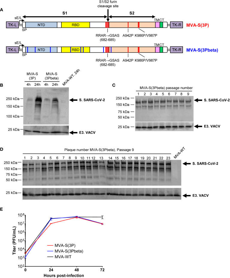
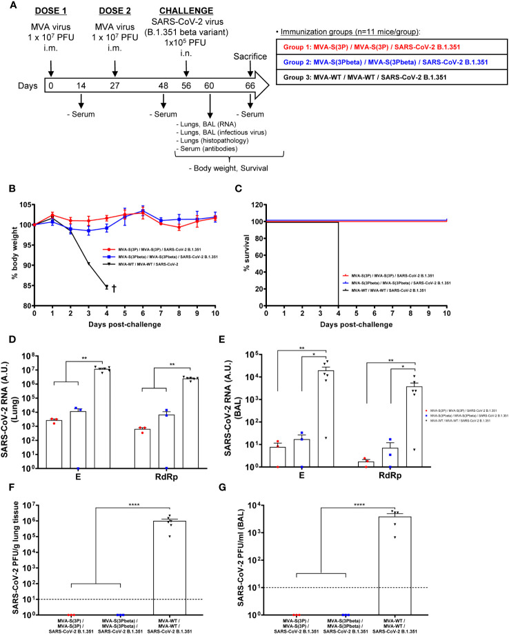
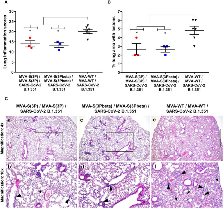
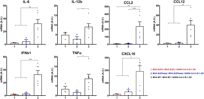
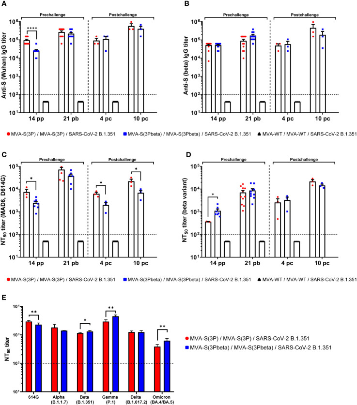
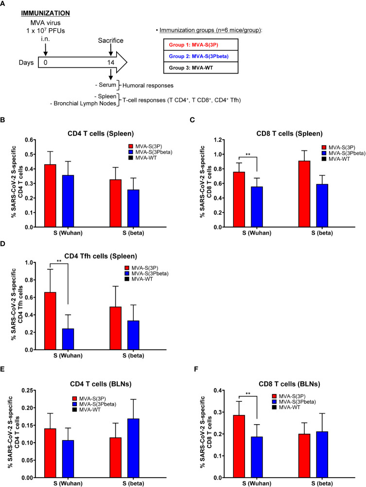
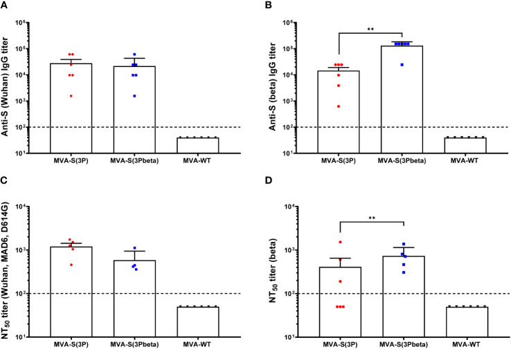
The constant appearance of new severe acute respiratory syndrome coronavirus 2 (SARS-CoV-2) variants of concern (VoCs) has jeopardized the protective capacity of approved vaccines against coronavirus disease-19 (COVID-19). For this reason, the generation of new vaccine candidates adapted to the emerging VoCs is of special importance. Here, we developed an optimized COVID-19 vaccine candidate using the modified vaccinia virus Ankara (MVA) vector to express a full-length prefusion-stabilized SARS-CoV-2 spike (S) protein, containing 3 proline (3P) substitutions in the S protein derived from the beta (B.1.351) variant, termed MVA-S(3Pbeta). Preclinical evaluation of MVA-S(3Pbeta) in head-to-head comparison to the previously generated MVA-S(3P) vaccine candidate, expressing a full-length prefusion-stabilized Wuhan S protein (with also 3P substitutions), demonstrated that two intramuscular doses of both vaccine candidates fully protected transgenic K18-hACE2 mice from a lethal challenge with SARS-CoV-2 beta variant, reducing mRNA and infectious viral loads in the lungs and in bronchoalveolar lavages, decreasing lung histopathological lesions and levels of proinflammatory cytokines in the lungs. Vaccination also elicited high titers of anti-S Th1-biased IgGs and neutralizing antibodies against ancestral SARS-CoV-2 Wuhan strain and VoCs alpha, beta, gamma, delta, and omicron. In addition, similar systemic and local SARS-CoV-2 S-specific CD4+ and CD8+ T-cell immune responses were elicited by both vaccine candidates after a single intranasal immunization in C57BL/6 mice. These preclinical data support clinical evaluation of MVA-S(3Pbeta) and MVA-S(3P), to explore whether they can diversify and potentially increase recognition and protection of SARS-CoV-2 VoCs.

Keywords: COVID-19; MVA-based vaccine; S protein; SARS-CoV-2; efficacy; immunogenicity; mice; variants of concern.


Conflict of interest statement

The authors declare that the research was conducted in the absence of any commercial or financial relationships that could be construed as a potential conflict of interest.

Figures

Figure 1
Figure 1
Design, generation, and in vitro characterization of MVA-S(3Pbeta) vaccine candidate. (A) Scheme of the prefusion-stabilized full-length S proteins inserted in the MVA genome to generate the MVA-S(3P) and MVA-S(3Pbeta) vaccine candidate. S1 and S2 regions are indicated, together with the amino acid mutations in the furin cleavage site and changes to prolines in the S2 region (indicated in red). The amino acid mutations of beta (B.1.351) variant are also indicated in blue. The SARS-CoV-2 S gene is inserted within the TK locus of MVA-WT virus and is driven by the sE/L virus promoter. NTD, N-terminal domain; RBD, receptor binding domain; TM, transmembrane; CT, cytoplasmic tail; TK-L, TK left; TK-R, TK right. (B) Expression of SARS-CoV-2 S protein by MVA-S(3P) and MVA-S(3Pbeta) vaccine candidates. Western blotting of MVA-infected (5 PFUs/cell) HeLa cell extracts at 4 and 24 hpi. Rabbit polyclonal anti-S and anti-VACV E3 antibodies were used for protein identification on 7% SDS-PAGE under reducing conditions. Size (in kilodaltons [kDa]) and migration of molecular weight markers are indicated. (C, D) Genetic stability of MVA-S(3Pbeta) vaccine candidate. Western blotting of DF-1 cell samples (24 hpi) infected with initial P2 stock and with 9 successive passages of MVA-S(3Pbeta) viruses (C) or from 23 individual virus plaques picked after 9 consecutive cell infection cycles (D). Samples were analyzed under reducing conditions. Rabbit polyclonal anti-S and anti-VACV E3 antibodies were used for protein identification. (E) Viral growth kinetics of MVA-S(3Pbeta). Monolayers of DF-1 cells were infected at 0.01 PFUs/cell with MVA-WT, MVA-S(3P) or MVA-S(3Pbeta). At different times postinfection (0, 24, 48, and 72 hpi), virus titers in cell lysates were quantified by a plaque immunostaining assay. The means of results from two independent experiments are shown.
Figure 2
Figure 2
MVA-S(3P) and MVA-S(3Pbeta) vaccine candidates protects K18-hACE2 transgenic mice from SARS-CoV-2 beta (B.1.351) infection. (A) Efficacy schedule. Female K18-hACE2 transgenic mice (n=11 per group) were immunized by the IM route with two doses, spanned by 4 weeks, of 1 x 10⁷ PFUs of MVA-S(3P), MVA-S(3Pbeta) or MVA-WT as indicated. At day 14 post-prime and 21 post-boost, serum samples were obtained from each mouse, as indicated. At day 56 (4 weeks post-boost) mice were challenged intranasally with 1 x 10⁵ PFUs of SARS-CoV-2 beta variant (B.1.351). At day 4 postchallenge, at least 3 mice per group were sacrificed and lungs, BAL and serum samples collected as indicated. Serum was also collected at day 10 postchallenge in groups 1, 2 (in group 3 all mice have died by this day). (B, C) The challenged mice were monitored for change of body weight (B) and mortality (C) for 10 days. †: mice were euthanized due to loss of more than 20% of initial body weight. (D, E) Virus replication in lung samples (D) and BAL (E). SARS-CoV-2 subgenomic E and genomic RdRp mRNA detected by RT-qPCR at 4 days after virus infection. Mean RNA levels (in arbitrary units [A.U.] normalized to uninfected mice) from duplicates of each lung and BAL samples and SEM of each group are represented. (F, G) SARS-CoV-2 infectious virus in lung samples (F) and BAL (G). Mean (PFUs/g of lung tissue or PFUs/mL of BAL) from triplicates of each sample and SEM of each group are represented. The dashed line represents the limit of detection. Ordinary one-way ANOVA of transformed data followed by Tukey’s multiple comparison test: *p < 0.033; **p < 0.002; ****p<0.0001.
Figure 3
Figure 3
MVA-S(3P) and MVA-S(3Pbeta) vaccine candidates reduced SARS-CoV-2 beta variant lung pathology in K18-hACE2 transgenic mice. (A) Mean and SEM of cumulative histopathological lesion scores in lung samples taken from immunized K18-hACE2 mice that were euthanized at day 4 post-challenge. Unpaired nonparametric Mann-Whitney test: *p < 0.033. (B) Percentage of lung area affected by inflammatory lesions in lung samples taken from immunized K18-hACE2 mice that were euthanized at day 4 post-challenge. Ordinary one-way ANOVA followed by Tukey’s multiple comparison test: *p < 0.033. (C) Representative lung histopathological sections (H&E staining) observed in immunized K18-hACE2 transgenic mice that were euthanized at day 4 post-challenge. A general view of the lung area (magnification: 4x) along with histopathological details from selected lung areas (black boxes) have been displayed (magnification: 10x). In mice that were immunized with two doses of MVA-S(3P) and MVA-S(3Pbeta) (a, c), alveolar spaces were larger and evident while inflammatory changes were less severe than those observed in mice immunized with two doses of MVA-WT (e). Mice immunized with MVA-S(3P) and MVA-S(3Pbeta) showed mild lung lesions that were characterized by the presence of small perivascular and peribronchiolar mononuclear infiltrates mainly constituted by lymphocytes (arrowheads), as well as occasional alveoli with some scarce infiltrated mononuclear cells (arrows) (b, d). On the contrary, in mice immunized with MVA-WT (control non-protected group) inflammatory lesions were more severe and diffuse. Such lesions were characterized by the presence of numerous large and perivascular and peribronchiolar infiltrates (arrowheads), alveolar spaces densely populated by inflammatory cells (arrows) and generalized alveolar septa thickening (f).
Figure 4
Figure 4
Vaccination with MVA-S(3P) or MVA-S(3Pbeta) diminished levels of proinflammatory cytokines in K18-hACE2 transgenic mice. mRNA levels of several cytokines/chemokines were detected by RT-qPCR in lungs obtained at 4 days postchallenge. Mean RNA levels (in A.U. normalized to uninfected mice) from duplicates of each sample and SEM of each group are represented. Ordinary one-way ANOVA of transformed data followed by Tukey’s multiple comparison test: *p < 0.033; **p < 0.002; ***p < 0.0002.
Figure 5
Figure 5
MVA-S(3P) and MVA-S(3Pbeta) vaccine candidates induced high levels of SARS-CoV-2-specific humoral immune responses in vaccinated and challenged K18-hACE2 transgenic mice. (A, B) Anti-S IgG titers against Wuhan (A) and beta variant (B) determined by ELISA in individual mouse serum samples from K18-hACE2 mice collected at day 14 post-prime (pp), 21 post-boost (pb), 4 and 10 postchallenge (pc). Mean endpoint titers of each sample from duplicates and SEM from each group are represented. Dashed line represents the limit of detection. Unpaired nonparametric Mann-Whitney test of transformed data: ****p < 0.0001. (C, D) SARS-CoV-2 neutralizing antibody titers against ancestral Wuhan strain (isolate MAD6, having D614G mutation) (C) and beta variant (B.1.351) (D). NT50 titers were evaluated in individual mouse serum samples collected at day 14 pp, 21 pb, 4 and 10 pc) using a live virus MNT assay. Mean NT50 values of each sample from triplicates and SEM from each group are represented. Dotted line represented the limit of detection. Unpaired t-test of transformed data: *p < 0.033. (E) SARS-CoV-2 neutralizing antibody titers against several SARS-CoV-2 VoCs. NT50 titers were evaluated in pooled mouse serum samples collected at day 14 pp and 21 pb, using VSV-based pseudoparticles expressing the SARS-CoV-2 S protein of different VoCs. Mean NT50 values and 95% confidence intervals from triplicates of each pooled group sample are represented. The dashed line represents the limit of detection. Unpaired t-test with Welch’s correction of transformed data: *p < 0.033; **p < 0.002.
Figure 6
Figure 6
SARS-CoV-2-specific immunogenicity in C57BL/6 mice immunized with one IN dose of MVA-S(3P) and MVA-S(3Pbeta) vaccine candidates. (A) Immunogenicity study schedule. Groups of female C57BL/6 mice (n=6 per group; 6 to 8 weeks old) were slightly anesthetized and each mouse received one dose of 1 × 10⁷ PFUs of MVA-S(3P), or MVA-S(3Pbeta) by the IN route in 50 μl of PBS. Mice inoculated with nonrecombinant MVA-WT were used as a control group. At day 14 after the immunization, mice were euthanized and blood, spleens and BLNs from each mouse were collected. (B–F) SARS-CoV-2 S-specific T-cellular immune responses were evaluated in spleens (B, C, D), and BLNs (E, F). Cell percentages were determined by ICS. (B, C) Magnitude of Wuhan strain and beta variant S-specific CD4+ (B) and CD8+ (C) T-cell immune responses in spleens. Percentages of CD4+ or CD8+ T cells expressing CD107a and/or producing IFN-γ and/or TNF-α and/or IL-2. (D) Magnitude of Wuhan strain and beta variant S-specific CD4+ Tfh cell responses in spleen. Percentages of CD4+ Tfh cells expressing CD40L and/or producing IFN-γ and/or IL-21. (E, F) Magnitude of Wuhan strain and beta variant S-specific CD4+ (E) and CD8+ (F) T-cell immune responses in BLNs. Percentages of CD4+ or CD8+ T cells expressing CD107a and/or producing IFN-γ and/or TNF-α and/or IL-2. **p < 0.002.
Figure 7
Figure 7
SARS-CoV-2-specific humoral immune responses elicited in C57BL/6 mice immunized with one IN dose of MVA-S(3P) and MVA-S(3Pbeta) vaccine candidates. SARS-CoV-2-specific humoral immune responses were evaluated in serum obtained at 14 days postimmunization. (A, B) Titers of binding IgG antibodies specific for the S protein from Wuhan strain (A) and beta variant (B), determined by ELISA in individual mouse serum samples in duplicate. Mean values and SEM are represented. The dashed line represents the limit of detection. Unpaired nonparametric Mann-Whitney test of transformed data: **p < 0.002. (C, D) SARS-CoV-2 NT50 antibody titers determined in individual mouse serum samples by using a live virus MNT assay. Mean NT50 values and SEM against parental Wuhan strain virus (MAD6 isolate, containing D614G mutation) (C) or beta variant (D) are represented. The dashed line represents the limit of detection. Unpaired t-test of transformed data.

Similar articles

Cited by

References

    1. Hillary VE, Ceasar SA. An update on COVID-19: SARS-CoV-2 variants, antiviral drugs, and vaccines. Heliyon (2023) 9(3):e13952. doi: 10.1016/j.heliyon.2023.e13952 - DOI - PMC - PubMed
    1. Dejnirattisai W, Zhou D, Supasa P, Liu C, Mentzer AJ, Ginn HM, et al. . Antibody evasion by the P.1 strain of SARS-CoV-2. Cell (2021) 184(11):2939–54:e9. doi: 10.1016/j.cell.2021.03.055 - DOI - PMC - PubMed
    1. Fernandes Q, Inchakalody VP, Merhi M, Mestiri S, Taib N, Moustafa Abo El-Ella D, et al. . Emerging COVID-19 variants and their impact on SARS-CoV-2 diagnosis, therapeutics and vaccines. Ann Med (2022) 54(1):524–40. doi: 10.1080/07853890.2022.2031274 - DOI - PMC - PubMed
    1. Garcia-Beltran WF, Lam EC, St Denis K, Nitido AD, Garcia ZH, Hauser BM, et al. . Multiple SARS-CoV-2 variants escape neutralization by vaccine-induced humoral immunity. Cell (2021) 184(9):2523. doi: 10.1016/j.cell.2021.04.006 - DOI - PMC - PubMed
    1. Moore JP, Offit PA. SARS-CoV-2 vaccines and the growing threat of viral variants. JAMA (2021) 325(9):821–2. doi: 10.1001/jama.2021.1114 - DOI - PubMed

Publication types

Supplementary concepts