Photoacoustic imaging plus X: a review
- PMID: 38156064
- PMCID: PMC10753847
- DOI: 10.1117/1.JBO.29.S1.S11513
Photoacoustic imaging plus X: a review
Abstract
Significance: Photoacoustic (PA) imaging (PAI) represents an emerging modality within the realm of biomedical imaging technology. It seamlessly blends the wealth of optical contrast with the remarkable depth of penetration offered by ultrasound. These distinctive features of PAI hold tremendous potential for various applications, including early cancer detection, functional imaging, hybrid imaging, monitoring ablation therapy, and providing guidance during surgical procedures. The synergy between PAI and other cutting-edge technologies not only enhances its capabilities but also propels it toward broader clinical applicability.
Aim: The integration of PAI with advanced technology for PA signal detection, signal processing, image reconstruction, hybrid imaging, and clinical applications has significantly bolstered the capabilities of PAI. This review endeavor contributes to a deeper comprehension of how the synergy between PAI and other advanced technologies can lead to improved applications.
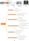
Approach: An examination of the evolving research frontiers in PAI, integrated with other advanced technologies, reveals six key categories named "PAI plus X." These categories encompass a range of topics, including but not limited to PAI plus treatment, PAI plus circuits design, PAI plus accurate positioning system, PAI plus fast scanning systems, PAI plus ultrasound sensors, PAI plus advanced laser sources, PAI plus deep learning, and PAI plus other imaging modalities.
Results: After conducting a comprehensive review of the existing literature and research on PAI integrated with other technologies, various proposals have emerged to advance the development of PAI plus X. These proposals aim to enhance system hardware, improve imaging quality, and address clinical challenges effectively.
Conclusions: The progression of innovative and sophisticated approaches within each category of PAI plus X is positioned to drive significant advancements in both the development of PAI technology and its clinical applications. Furthermore, PAI not only has the potential to integrate with the above-mentioned technologies but also to broaden its applications even further.
Keywords: circuit design; deep learning; laser source; multimodal imaging; photoacoustic imaging; treatment; ultrasound sensor.
© 2023 The Authors.
Figures


Similar articles
-
Prescription of Controlled Substances: Benefits and Risks.2025 Jul 6. In: StatPearls [Internet]. Treasure Island (FL): StatPearls Publishing; 2025 Jan–. 2025 Jul 6. In: StatPearls [Internet]. Treasure Island (FL): StatPearls Publishing; 2025 Jan–. PMID: 30726003 Free Books & Documents.
-
Short-Term Memory Impairment.2024 Jun 8. In: StatPearls [Internet]. Treasure Island (FL): StatPearls Publishing; 2025 Jan–. 2024 Jun 8. In: StatPearls [Internet]. Treasure Island (FL): StatPearls Publishing; 2025 Jan–. PMID: 31424720 Free Books & Documents.
-
Management of urinary stones by experts in stone disease (ESD 2025).Arch Ital Urol Androl. 2025 Jun 30;97(2):14085. doi: 10.4081/aiua.2025.14085. Epub 2025 Jun 30. Arch Ital Urol Androl. 2025. PMID: 40583613 Review.
-
Photoacoustic imaging for cutaneous melanoma assessment: a comprehensive review.J Biomed Opt. 2024 Jan;29(Suppl 1):S11518. doi: 10.1117/1.JBO.29.S1.S11518. Epub 2024 Jan 12. J Biomed Opt. 2024. PMID: 38223680 Free PMC article.
-
Revolutionizing the Pancreatic Tumor Diagnosis: Emerging Trends in Imaging Technologies: A Systematic Review.Medicina (Kaunas). 2024 Apr 24;60(5):695. doi: 10.3390/medicina60050695. Medicina (Kaunas). 2024. PMID: 38792878 Free PMC article.
Cited by
-
Nanoporous Submicron Gold Particles Enable Nanoparticle-Based Localization Optoacoustic Tomography (nanoLOT).Small. 2024 Dec;20(51):e2404904. doi: 10.1002/smll.202404904. Epub 2024 Oct 12. Small. 2024. PMID: 39394978 Free PMC article.
-
Iterative optimization algorithm with structural prior for artifacts removal of photoacoustic imaging.Photoacoustics. 2025 Apr 26;44:100726. doi: 10.1016/j.pacs.2025.100726. eCollection 2025 Aug. Photoacoustics. 2025. PMID: 40454259 Free PMC article.
-
Multiparametric Brain Hemodynamics Imaging Using a Combined Ultrafast Ultrasound and Photoacoustic System.Adv Sci (Weinh). 2024 Aug;11(31):e2401467. doi: 10.1002/advs.202401467. Epub 2024 Jun 17. Adv Sci (Weinh). 2024. PMID: 38884161 Free PMC article.
-
Signal-domain speed-of-sound correction for ring-array-based photoacoustic tomography.Photoacoustics. 2025 May 22;44:100735. doi: 10.1016/j.pacs.2025.100735. eCollection 2025 Aug. Photoacoustics. 2025. PMID: 40502803 Free PMC article.
-
Adaptive Vectorial Restoration from Dynamic Speckle Patterns Through Biological Scattering Media Based on Deep Learning.Sensors (Basel). 2025 Mar 14;25(6):1803. doi: 10.3390/s25061803. Sensors (Basel). 2025. PMID: 40292951 Free PMC article.
References
-
- Wang L. V., “Tutorial on photoacoustic microscopy and computed tomography,” IEEE J. Sel. Top. Quantum Electron. 14(1), 171–179 (2008).IJSQEN10.1109/JSTQE.2007.913398 - DOI
-
- Basij M., et al. , “Development of an integrated photoacoustic-guided laser ablation intracardiac theranostic system,” in IEEE Int. Ultrasonics Symp. (IUS), pp. 1–4 (2021).10.1109/IUS52206.2021.9593442 - DOI
-
- Gao S., et al. , “Photoacoustic necrotic region mapping for radiofrequency ablation guidance,” in IEEE Int. Ultrasonics Symp. (IUS), pp. 1–4 (2021).10.1109/IUS52206.2021.9593388 - DOI

