. 2024 Feb 1;32(2):131-147.e7.
doi: 10.1016/j.str.2023.11.015. Epub 2023 Dec 28.

Antibody targeting of conserved sites of vulnerability on the SARS-CoV-2 spike receptor-binding domain

Affiliations

Antibody targeting of conserved sites of vulnerability on the SARS-CoV-2 spike receptor-binding domain

Rajeshwer S Sankhala et al. Structure. .

Abstract

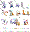
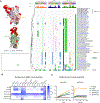
Given the continuous emergence of severe acute respiratory syndrome coronavirus 2 (SARS-CoV-2) variants of concern (VoCs), immunotherapeutics that target conserved epitopes on the spike (S) glycoprotein have therapeutic advantages. Here, we report the crystal structure of the SARS-CoV-2 S receptor-binding domain (RBD) at 1.95 Å and describe flexibility and distinct conformations of the angiotensin-converting enzyme 2 (ACE2)-binding site. We identify a set of SARS-CoV-2-reactive monoclonal antibodies (mAbs) with broad RBD cross-reactivity including SARS-CoV-2 Omicron subvariants, SARS-CoV-1, and other sarbecoviruses and determine the crystal structures of mAb-RBD complexes with Ab246 and CR3022 mAbs targeting the class IV site, WRAIR-2134, which binds the recently designated class V epitope, and WRAIR-2123, the class I ACE2-binding site. The broad reactivity of class IV and V mAbs to conserved regions of SARS-CoV-2 VoCs and other sarbecovirus provides a framework for long-term immunotherapeutic development strategies.


Conflict of interest statement

Declaration of interests Patent application number PCT/US 63/140,763 was filed containing mAbs described in this publication for authors S.J.K., K.M., V.D., S.M.T., G.D., and N.L.M. The status of the patent is pending, not yet published. M.G.J. and K.M. are named as inventors on International Patent Application No. WO 2021/178971 A1 entitled “Vaccines against SARS-CoV-2 and other coronaviruses.” M.G.J. is named as an inventor on International Patent Application No. WO/2018/081318 and U.S. patent 10,960,070 entitled “Prefusion Coronavirus S Proteins and Their Use.”

Figures
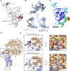
Figure 1.
Figure 1.. Crystal structure of the SARS-CoV-2 Receptor Binding Domain (RBD).
A, The SARS-CoV-2 RBD is shown in ribbon representation, glycan N343 is shown in sphere representation, with the N- and C-termini shown as green and red spheres, respectively. Two ACE2 ridge loop conformations are labeled and colored white and brown. B, Top panel, 2Fo-Fc electron density of the ACE2 binding loop, contoured at 1 σ. Bottom panel, The ACE2 binding loop, with either Forms-1 or −2 modeled into the corresponding density. C, SARS-CoV-2 RBD is shown as a ribbon cartoon colored by the temperature factor value (B factor) in a gradient from blue to red, spanning a numeric B-factor range from 6.5 to 96.5. D, The SARS-CoV-2 RBD structure (white) is overlaid with the ACE2-bound structure of SARS-CoV-2 RBD (blue) (PDB code: 6M0J). E, Top panel, zoom-in panel showing close contacts of ACE2 residues with Form-1 conformation of RBD. Bottom panel, zoom-in panel showing close contacts of ACE2 residues with Form-2 conformation.
Figure 2.
Figure 2.. Characterization of SARS-CoV-2 RBD-targeted mAbs.
A, Epitope binning of RBD-directed mAbs via a BLI-based competition assay. Values represent the % residual binding of the indicated second antibody after saturation of the antigen (RBD molecule) with the indicated first antibody. Shading from dark to light red indicates competition strength ranging from strong (0–25%), to lack thereof (>50%). Competition groups are indicated by black boxes. B, Binding of mAbs to VoC and VoI RBD mutants assessed by BLI. Heat-map shows the fold change in binding responses relative to the WA-1 RBD protein with loss and gain in binding represented in shades of red and blue, respectively. C, Neutralization activity of RBD mAbs against a panel of pseudotyped viruses representing the VOCs and SARS-CoV-1. Heat-map indicates IC50 values (μg/mL) ranging from potently neutralizing (blue), to poorly neutralizing (dark red). D, Assessment of RBD mAbs recruitment of Fc-mediated opsonization, antibody dependent complement deposition (ADCD), antibody dependent cell cytotoxicity (ADCC) and trogocytosis. R10 is a media-only control. E, ACE2 blocking activity of RBD mAbs. mAbs were assessed for their ability to block ACE2 binding to WA-1 (left) and Omicron (right) RBDs in a BLI-based assay.
Figure 3.
Figure 3.. Crystal structure of WRAIR-2134 antibody in complex with SARS-CoV-2 RBD.
A, Left, crystal structure of WRAIR-2134, in complex with SARS-CoV-2 RBD. Both the RBD and WRAIR-2134 are shown in cartoon representation. RBD is colored white, while heavy and light chains of WRAIR-2134 are colored dark and light green, respectively. ACE2 binding site on the RBD is colored dark gray. Right, Epitope footprint of WRAIR-2134 is shown on the surface of the RBD and colored based on the antibody heavy and light chain colors. ACE2 epitope is highlighted in dark gray. B, Left, SARS-CoV-2 RBD is shown in surface representation with WRAIR-2134 epitope highlighted in green. Contacting residues from RBD are shown in stick representation and contributing heavy and light chain CDRs are shown and labelled. Right, buried surface area (BSA) for the heavy and light chain CDR loops are shown as a bar diagram. C, Crystal structures of WRAIR-2057-RBD and S2H97-RBD complexes are overlaid on the WRAIR-2134-RBD structure. The WRAIR-2134 epitope is colored dark green on the surface of SARS-CoV-2 RBD while WRAIR-2057 and S2H97 are shown in cartoon representation. Side and top views are shown. All three antibodies target an overlapping epitope while approaching the RBD at slightly different angles. D, WRAIR-2057 (magenta) and S2H97 (teal) epitopes are shown on the surface of SARS-CoV-2 RBD with ACE2 epitope highlighted in dark gray color. WRAIR-2134 and ION-300 epitopes are outlined in dark green and light blue colors, respectively. E, Omicron mutations are highlighted as red spheres on the surface of SARS-CoV-2 RBD. WRAIR-2134 epitope is shown in the cartoon tube representation and colored dark green. F, Sequence alignment of SARS-CoV-2 RBD with WRAIR-2134, WRAIR-2057, S2H97, ION-300 and ACE2 epitope residues highlighted in dark green, magenta, teal, blue, and gray, respectively.
Figure 4.
Figure 4.. Crystal structure of WRAIR-2123 antibody in complex with SARS-CoV-2 RBD.
A, Left, crystal structure of WRAIR-2123, in complex with SARS-CoV-2 RBD. Both the RBD and WRAIR-2123 are shown in cartoon representation. RBD is colored white, while heavy and light chains of WRAIR-2123 are shown in dark and light blue colors, respectively. ACE-2 binding site on the RBD is colored dark gray. Right, Epitope footprint of WRAIR-2123 are shown on the surface of the RBD and colored based on the antibody heavy and light chain colors. ACE2 epitope is indicated by a dark gray line. B, SARS-CoV-2 RBD is shown in surface representation with WRAIR-2123 epitope highlighted in blue. Contacting residues from the RBD are shown in stick representation and heavy and light chain contacting CDRs are shown in ribbon representation. C, Buried surface area (BSA) for the heavy and light chain CDR loops are shown as a bar diagram. D, Omicron mutations are highlighted as red spheres on the surface of SARS-CoV-2 RBD. The WRAIR-2123 epitope is shown in tubular representation and colored blue. Omicron mutations that fall in the mAb epitope are colored blue and labeled. E, Sequence alignment of SARS-CoV-2 RBD with WRAIR-2123 and ACE2 epitope residues highlighted in blue and gray, respectively.
Figure 5.
Figure 5.. Crystal structures of antibodies CR3022 and Ab246 in complex with SARS-CoV-2 RBD.
A, Left, crystal structure of Ab246, in complex with SARS-CoV-2 RBD. Both the RBD and Ab246 are shown in cartoon representation. RBD is colored white, while heavy and light chains of Ab246 are shown in dark blue and light blue colors, respectively. ACE2 binding site on the RBD is colored dark gray. Right, Epitope footprint of Ab246 is shown on the surface of the RBD and colored based on the antibody heavy and light chain colors. ACE2 epitope is indicated by a dark gray line. B, Left, SARS-CoV-2 RBD is shown in surface representation with Ab246 epitope highlighted in blue. Contacting residues from RBD are shown in stick representation and contributing heavy and light chain CDRs are shown and labelled. CDR loops are designated using the Kabat numbering system. Right, buried surface area (BSA) for the heavy and light chain CDR loops are shown as bar diagram. C, Left, crystal structure of CR3022 in complex with SARS-CoV-2 RBD. Both the RBD and CR3022 are shown in cartoon representation. RBD is colored white, while heavy and light chains of CR3022 are shown in dark and light orange colors, respectively. ACE2 binding site on the RBD is colored dark gray. Right, Epitope footprint of CR3022 is shown on the surface of the RBD and colored based on the antibody heavy and light chain colors. ACE2 epitope is indicated by a dark gray line. D, Left, SARS-CoV-2 RBD is shown in surface representation with CR3022 epitope highlighted in orange. Contacting residues from RBD are shown in stick representation and CDR contact loops are shown and labelled. CDR loops were assigned using the Kabat system. Right, buried surface area (BSA) for the heavy and light chain CDR loops are shown as a bar diagram. E, Binding interface of Ab246 CDR H3 loop and RBD is shown. CDR H3 is shown as dark blue ribbon with disulfide bond displayed in stick representation. F, Crystal structure of CR3022-RBD complex is overlaid with the Ab246-RBD structure. Both antibodies target an overlapping epitope while approaching at a right angle to each other. G, Omicron mutations are highlighted as red spheres on the SARS-CoV-2 RBD. CR3022 and Ab246 epitopes are shown in tubular representation and colored orange and dark blue, respectively, with overlapping regions of the two epitopes colored dark brown. The single Omicron mutated residue S375 that is in the CR3022 epitope is colored orange. H, Sequence alignment of SARS-CoV-2 RBD with CR3022 and Ab246 epitopes highlighted in orange and blue, respectively. ACE-2 epitope is highlighted in gray. Pro384, mutated to Asn, is highlighted in red.
Figure 6.
Figure 6.. mAb class assignment and epitope accessibility.
A, Structures of WRAIR-2134-RBD, WRAIR-2057-RBD and S2H97-RBD (PDB code: 7M7W) complexes are overlaid with previously reported antibodies that represent frequently observed SARS-CoV-2 antibody classes. RBD and representative mAbs are shown in surface representation. mAbs WRAIR-2057 and S2H97 are shown as ribbon and cartoon representation in magenta and teal colors, respectively. WRAIR-2134 epitope is shown in dark green color on the RBD surface. B, Epitopes for WRAIR-2057, S2H97 and ION-300 are outlined and colored magenta, teal and blue, respectively. C, Structural alignment of the WRAIR-2134-RBD and WRAIR-2057-RBD complexes with closed conformation of SARS-CoV-2 S-2P (all RBD down conformation, PDB code: 6ZGE). One protomer of the S trimer is displayed in surface representation, with the RBD of interest labeled, and epitopes for the WRAIR-2134 and WRAIR-2057 are shown on the surface of RBD, from a single protomer, in dark green and magenta colors, respectively. The other two protomers are shown as cartoon representation. In this overlay, the Fv region of WRAIR-2134 and both the Fv and Fc1 regions of WRAIR-2057 clash with the NTD of a neighboring protomer. D, Structure alignment of the WRAIR-2134-RBD and WRAIR-2057-RBD complexes with open conformation of SARS-CoV-2 S-2P (1-RBD up conformation, PDB code: 7BWJ). In this overlay, both Fab molecules have full access to the RBD, with no clashes observed.
Figure 7.
Figure 7.. Epitope conservation and mAb cross-reactivity analysis.
A, (Top) Structural and sequence analysis of the CR3022, Ab246 and WRAIR-2134 footprints across sarbecoviruses. The epitope residues are numbered according to the Wuhan reference; the strength of the interaction between the mAb and the RBD is indicated by the height and color of the histogram bars above the sequence alignment. Sequences are ordered based on their phylogenetic relationships based on a maximum likelihood phylogenetic tree derived from RBD protein sequences. (Left) The RBD structure is shown in surface representation and depicts mutations between SARS-CoV-1 and SARS-CoV-2 in red; the CR3022, Ab246, and WRAIR-2134 epitopes are outlined and labeled. B, mAb binding to sarbecovirus RBDs using BLI to assess cross-reactivity. Heat-map shows the binding responses (nm) represented in shades of purple. C, Cross-reactivity of mAbs were also assessed for binding to bat SARS-related CoV Rs4874 and Rs4231 S glycoproteins, using BLI.

References

    1. Munster VJ, Koopmans M, van Doremalen N, van Riel D, and de Wit E (2020). A Novel Coronavirus Emerging in China - Key Questions for Impact Assessment. The New England journal of medicine 382, 692–694. 10.1056/NEJMp2000929. - DOI - PubMed
    1. Beaudoin-Bussieres G, Laumaea A, Anand SP, Prevost J, Gasser R, Goyette G, Medjahed H, Perreault J, Tremblay T, Lewin A, et al. (2020). Decline of Humoral Responses against SARS-CoV-2 Spike in Convalescent Individuals. mBio 11. 10.1128/mBio.02590-20. - DOI - PMC - PubMed
    1. Kustin T, Harel N, Finkel U, Perchik S, Harari S, Tahor M, Caspi I, Levy R, Leshchinsky M, Ken Dror S, et al. (2021). Evidence for increased breakthrough rates of SARS-CoV-2 variants of concern in BNT162b2-mRNA-vaccinated individuals. Nature medicine 27, 1379–1384. 10.1038/s41591-021-01413-7. - DOI - PMC - PubMed
    1. Long QX, Tang XJ, Shi QL, Li Q, Deng HJ, Yuan J, Hu JL, Xu W, Zhang Y, Lv FJ, et al. (2020). Clinical and immunological assessment of asymptomatic SARS-CoV-2 infections. Nature medicine 26, 1200–1204. 10.1038/s41591-020-0965-6. - DOI - PubMed
    1. Becker M, Dulovic A, Junker D, Ruetalo N, Kaiser PD, Pinilla YT, Heinzel C, Haering J, Traenkle B, Wagner TR, et al. (2021). Immune response to SARS-CoV-2 variants of concern in vaccinated individuals. Nature communications 12, 3109. 10.1038/s41467-021-23473-6. - DOI - PMC - PubMed

Publication types

LinkOut - more resources