. 2024 Aug;154(2):480-491.
doi: 10.1016/j.jaci.2023.11.923. Epub 2023 Dec 28.

Thymic stromal lymphopoietin induces IL-4/IL-13 from T cells to promote sebum secretion and adipose loss

Affiliations

Thymic stromal lymphopoietin induces IL-4/IL-13 from T cells to promote sebum secretion and adipose loss

Ruth Choa et al. J Allergy Clin Immunol. 2024 Aug.

Abstract

Background: The cytokine TSLP promotes type 2 immune responses and can induce adipose loss by stimulating lipid loss from the skin through sebum secretion by sebaceous glands, which enhances the skin barrier. However, the mechanism by which TSLP upregulates sebaceous gland function is unknown.

Objectives: This study investigated the mechanism by which TSLP stimulates sebum secretion and adipose loss.

Methods: RNA-sequencing analysis was performed on sebaceous glands isolated by laser capture microdissection and single-cell RNA-sequencing analysis was performed on sorted skin T cells. Sebocyte function was analyzed by histological analysis and sebum secretion in vivo and by measuring lipogenesis and proliferation in vitro.

Results: This study found that TSLP sequentially stimulated the expression of lipogenesis genes followed by cell death genes in sebaceous glands to induce holocrine secretion of sebum. TSLP did not affect sebaceous gland activity directly. Rather, single-cell RNA-sequencing revealed that TSLP recruited distinct T-cell clusters that produce IL-4 and IL-13, which were necessary for TSLP-induced adipose loss and sebum secretion. Moreover, IL-13 was sufficient to cause sebum secretion and adipose loss in vivo and to induce lipogenesis and proliferation of a human sebocyte cell line in vitro.

Conclusions: This study proposes that TSLP stimulates T cells to deliver IL-4 and IL-13 to sebaceous glands, which enhances sebaceous gland function, turnover, and subsequent adipose loss.

Keywords: IL-13; IL-4; Sebaceous gland; T cells; TSLP; adipose loss; sebum.


Conflict of interest statement

Conflict of Interest

U.S. Provisional Patent Application No. 62/972,462 was filed by the University of Pennsylvania. The inventors are Taku Kambayashi and Ruth Choa. The patent application is based on the finding that TSLP induces sebum secretion from the skin and that this can lead to treatment of skin conditions and obesity and its complications.

Figures

Fig 1.
Fig 1.
scRNAseq of skin T cells in mice given Ctrl-AAV vs. TSLP-AAV (A) Schematic of scRNAseq of skin T cells. Skin T cells were isolated and pooled from mice treated with Ctrl-AAV (N = 30) or TSLP-AAV (N = 10 mice). (B) TCR clonotype abundance and distribution from TCR sequencing of skin T cells. (C) UMAP of α or β TCR -expressing single cells with annotations. (D) UMAP plot comparison of α or β TCR -expressing single cells from mice treated with control vs. TSLP-AAV. (E) Heatmap analysis of the top 10 most differentially expressed genes per cluster. (F) GSEA analysis on the DEGs between activated CD4+ T cells (0, 1, 2, 3, 8, 9, and 13) in the TSLP condition and the Ctrl condition. (G) Targeted analysis and dot plot of Th2 genes.
Fig 2.
Fig 2.
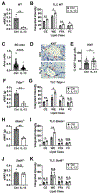
T cell derived IL-4 and 13 are necessary for TSLP-induced adipose loss and sebum secretion (A) eWAT masses and (B) TLC quantification of lipids extracted from hair of Stat6−/− mice, 2 weeks post AAV (N = 4-5 mice/group, 1 representative of 3 experiments). CE = cholesterol esters, WE = wax esters, FFA = fatty acids, and FC = free cholesterol. (C) eWAT masses and (D) TLC quantification of lipids extracted from hair of Il4/13fl/fl and VavCreIl4/13fl/fl mice, 2 weeks post AAV (N = 3 mice/group, 1 representative of 3 experiments). (E) eWAT masses and (F) TLC quantification of hair lipids from Rag2−/− mice transferred with T cells from either of Il4/13fl/fl or VavCreIl4/13fl/fl mice, 2 weeks post AAV (N = 3mice/group, 1 representative of 2 experiments). N.S. = not significant, *P<0.05, **P<0.01, ***P<0.001, ****P0.0001 by Student t-test. Data are shown as mean ± SEM.
Fig 3.
Fig 3.
IL-13 is sufficient to drive adipose loss and sebum hypersecretion. (A) eWAT masses and (B) TLC quantification of hair lipids from wildtype (WT) mice, 2 weeks post AAV (N = 3 mice/group). (C) Quantification of sebaceous gland (SG) area in WT mice, 2 weeks post AAV (n = 105 Ctrl and 95 TSLP SGs from 3 mice/group). (D) Ki67 staining (red arrows indicate Ki67+ cells) and (E) quantification of SGs in WT mice, 2 weeks post AAV (N = 3 mice/group). (F) eWAT masses and (G) TLC quantification of hair lipids from Tslpr−/− mice, 2 weeks post AAV (N = 3 mice/group). (H) eWAT masses and (I) TLC quantification of hair lipids from Ebeta−/− mice, 2 weeks post AAV (N = 3 mice/group). t-test. (J) eWAT masses and (K) TLC quantification of hair lipids from Stat6−/− mice, 2 weeks post AAV (N = 4 mice/group). For each experiment, 1 representative of 2 experiments is shown. N.S. = not significant, *P<0.05, **P<0.01, ****P<0.0001 by Student t-test. Data are shown as mean ± SEM.
Fig 4.
Fig 4.
IL-4 and 13 induce lipogenesis and proliferation of SEB-1 cells. (A) LipidTOX flow staining of SEB-1 cells treated with media alone, media with insulin, or media with IL-4 and 13 (N = 3 samples/group, 1 representative of 2 experiments). MFI = mean fluorescence intensity. (B) TLC triglyceride (TG) levels of SEB-1 cells treated with or without IL-4 and 13 (N = 6 samples/group, 1 representative of 3 experiments). (C) Crystal violet optical density (OD) time course over 5 days of treatment with or without IL-4 and 13 (N = 5 samples/group, 1 representative of 3 experiments). N.S. = not significant, *P<0.05, **P<0.01, ***P<0.001, ****P<0.0001 by Student t-test. Data are shown as mean ± SEM.
Fig 5.
Fig 5.
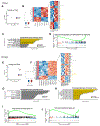
TSLP promotes lipogenesis at day 7 and cell death at day 14 in sebaceous glands in vivo. LCM of isolated skin sebaceous glands and RNAseq analysis at 7 (Fig 5, A–D, n = 3 samples/group) and 14 (Fig 5, E–J, n = 5 samples/group) days after control vs. TSLP-AAV injection. (A) Principle component analysis (PCA) of control (blue) vs. TSLP (red) samples. (B) Heat map of DEGs, zoomed in on cell death and lipid metabolism genes. (C) Upregulated GO terms induced by TSLP. (D) GSEA plot representing a downregulated pathway induced by TSLP, identified via the C2 curated MSigDB database. (E) PCA of control (blue) vs. TSLP (red) samples. (F) Heat map of DEGs, zoomed in on cell death and lipid metabolism genes. (G) Upregulated GO terms induced by TSLP. (H) Downregulated GO terms induced by TSLP. (I) GSEA representing enrichment of cell death genes, identified via the Reactome database. (J) GSEA representing downregulation of fatty acid metabolism genes with TSLP, identified via the KEGG database. All GO terms were identified from the GO knowledgebase; FDR adj-p-val < 0.05; number listed beside each bar corresponds to number of DEGs overlapping with genes in term dataset.

References

    1. Eyerich S, Eyerich K, Traidl-Hoffmann C, Biedermann T. Cutaneous Barriers and Skin Immunity: Differentiating A Connected Network. Trends Immunol. 2018;39(4):315–27. - PubMed
    1. Picardo M, Mastrofrancesco A, Bíró T. Sebaceous gland-a major player in skin homoeostasis. Exp Dermatol. 2015;24(7):485–6. - PubMed
    1. Lee DY, Yamasaki K, Rudsil J, Zouboulis CC, Park GT, Yang JM, et al. Sebocytes express functional cathelicidin antimicrobial peptides and can act to kill propionibacterium acnes. J Invest Dermatol. 2008;128(7):1863–6. - PMC - PubMed
    1. Nakatsuji T, Kao MC, Zhang L, Zouboulis CC, Gallo RL, Huang CM. Sebum free fatty acids enhance the innate immune defense of human sebocytes by upregulating beta-defensin-2 expression. J Invest Dermatol. 2010;130(4):985–94. - PMC - PubMed
    1. Pan Y, Tian T, Park CO, Lofftus SY, Mei S, Liu X, et al. Survival of tissue-resident memory T cells requires exogenous lipid uptake and metabolism. Nature. 2017;543(7644):252–6. - PMC - PubMed