. 2024 Feb;28(3):e18098.
doi: 10.1111/jcmm.18098. Epub 2023 Dec 30.

circELP2 reverse-splicing biogenesis and function as a pro-fibrogenic factor by targeting mitochondrial quality control pathway

Affiliations

circELP2 reverse-splicing biogenesis and function as a pro-fibrogenic factor by targeting mitochondrial quality control pathway

Songzi Zhang et al. J Cell Mol Med. 2024 Feb.

Abstract

Idiopathic pulmonary fibrosis (IPF) is considered as a chronic, fibrosing interstitial pneumonia with unknown mechanism. The present work aimed to explore the function, biogenesis and regulatory mechanism of circELP2 in pulmonary fibrosis and evaluate the value of blocking circELP2-medicated signal pathway for IPF treatment. The results showed that heterogeneous nuclear ribonucleoprotein L initiated reverse splicing of circELP2 resulting in the increase of circELP2 generation. The biogenetic circELP2 activated the abnormal proliferation and migration of fibroblast and extracellular matrix deposition to promote pulmonary fibrogenesis. Mechanistic studies demonstrated that cytoplasmic circELP2 sponged miR-630 to increase transcriptional co-activators Yes-associated protein 1 (YAP1) and transcriptional co-activator with PDZ-binding motif (TAZ). Then, YAP1/TAZ bound to the promoter regions of their target genes, such as mTOR, Raptor and mLST8, which in turn activated or inhibited the genes expression in mitochondrial quality control pathway. Finally, the overexpressed circELP2 and miR-630 mimic were packaged into adenovirus vector for spraying into the mice lung to evaluate therapeutic effect of blocking circELP2-miR-630-YAP1/TAZ-mitochondrial quality control pathway in vivo. In conclusion, blocking circELP2-medicated pathway can alleviate pulmonary fibrosis, and circELP2 may be a potential target to treat lung fibrosis.

Keywords: YAP1/TAZ; circRNA; miRNA; mitochondrial quality control pathway; pulmonary fibrosis.


Conflict of interest statement

The authors declared no potential conflicts of interest with respect to the research, authorship and/or publication of this article.

Figures
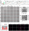
FIGURE 1
FIGURE 1
Cytoplasmic circELP2 accelerated fibroblast activation and matrix deposition in TGFβ1‐treated MRC‐5 cells. (A) qRT‐PCR detected that circELP2 increased significantly with time dependence in the TGFβ1‐stimulated group, reaching maximum value in 72 h. (B) The xCELLigence RTCA DPlus instrument monitored that knockdown circELP2 repressed activated fibroblast proliferation and migration. But overexpressed circELP2 enhanced the proliferation and migration. (C) The Incucyte S3 live‐cell analysis system automatically recorded the cell migration and exhibited that the activated fibroblast migration was blocked by circELP2 knockdown and promoted by circELP2 overexpression. (D) Western blot uncovered that the fibrotic and differentiation‐related proteins were inhibited by knockdown circELP2 and promoted by overexpressed circELP2. (E) Nuclear (grey) cytoplasmic (red) separation result manifested circELP2 mainly located in the cytoplasm. β‐Actin served as a cytoplasmic control. U6 served as a nuclear control. (F) RNA FISH images depicted that circELP2 (red) mainly located in the cytoplasm. Silencing circELP2 did not induce circELP2 translocation. 18S and U6 RNA were used as cytoplasmic and nuclear localization markers, respectively. DNA (blue) was stained with DAPI. si‐circELP2 NC indicates si‐circELP2 negative control. BP indicates blank plasmid, and RP indicates the recombinant plasmid of the overexpressed circELP2. Each bar represents mean ± SD (n = 6), *p < 0.05.
FIGURE 2
FIGURE 2
The hnRNP L initiated circELP2 back‐splicing generation. (A) Schematic illustration showing circELP2 produced from its host gene ELP2. Sanger sequencing after PCR with specified primers illustrated the circular junction of circELP2, and its full length was 1100 nt. (B) Agarose gel electrophoresis result revealed that circELP2 generated from cDNA, not gDNA. (C) After RNase R action, circELP2 was detected, but GAPDH and ELP2 were not detected. (D) The actinomycin D experiment indicated that circELP2 was more stable than ELP2. (E) qRT‐PCR analysed that hnRNP L was detectable in the anti‐hnRNP L RIP, but not undetectable in the IgG RIP. (F) si‐hnRNP L inhibited circELP2 and overexpressed hnRNP L promoted circELP2. (G) Dual‐luciferase experiment discovered that si‐hnRNP L repressed circELP2 fluorescence intensity. (H) Immunofluorescence result depicted that silencing hnRNP L made the cellular morphology normal and α‐SMA decreased, but overexpressed hnRNP L worsened the cellular state and α‐SMA increased. (I) The myofibroblast migration was inhibited by si‐hnRNP L and enhanced by overexpressed hnRNP L. (J) circELP2 overexpression reversed the downtrends of fibrotic and differentiation‐related proteins caused by interfering with hnRNP L. si‐circELP2 reversed the uptrends of fibrotic and differentiation‐related proteins induced by hnRNP L overexpression. Each bar represents mean ± SD (n = 6), *p < 0.05.
FIGURE 3
FIGURE 3
The circELP2 acted as a sponge of miR‐630 to promote pulmonary fibrosis. (A) Firefly and Renilla assay investigated that miR‐630 mimic markedly inhibited the luciferase activity. When the predicted binding site of circELP2 was mutated, miR‐630 mimic did not cause the change of luciferase activity. (B) RAP experiment proved that miR‐630 directly was binded to circELP2. (C) qRT‐PCR detection illustrated that miR‐630 expression was significantly reduced in MRC‐5 cells induced by 5 ng/mL TGFβ1 for 24, 48 and 72 h. (D) miR‐630 inhibitor obviously promoted vimentin, α‐SMA, collagen I and III, and miR‐630 mimic obviously inhibited their expressions. (E) Rescue experiment of Western blot exhibited that miR‐630 inhibitor reversed the downtrend of these fibrotic proteins induced by knockdown circELP2 and miR‐630 mimic reversed the uptrend of these proteins induced by overexpressed circELP2. (F) The proliferation and migration slowed down after miR‐630 mimic transfected into cell samples, but increased after miR‐630 inhibitor transfection. (G) Rescue experiments of proliferation and migration indicated that miR‐630 inhibitor reversed the downtrend of proliferation and migration induced by circELP2 knockdown and miR‐630 mimic reversed the uptrend of proliferation and migration induced by circELP2 overexpression. Each bar represents mean ± SD (n = 6), *p < 0.05.
FIGURE 4
FIGURE 4
The circELP2 targeted YAP1 and TAZ by sponging of miR‐630 to promote pulmonary fibrosis. (A) Firefly and Renilla dual‐luciferase test indicated that miR‐630 mimic obviously inhibited luciferase activity. When the predicted binding site of YAP1 and TAZ was mutated, miR‐630 mimic did not cause the change of luciferase activity. (B) Western blot elucidated that YAP1 and TAZ obviously increased in MRC‐5 cells induced by 5 ng/mL TGFβ1 at 24, 48 and 72 h. (C) Western blot demonstrated that miR‐630 mimic repressed YAP1 and TAZ expression and miR‐630 inhibitor increased their expression. (D) Rescue experiment presented that overexpressed YAP1 and TAZ rescued the downtrend of the fibrotic proteins induced by miR‐630 mimic. Silencing YAP1 and TAZ rescued the increasing trend of these proteins induced by miR‐630 inhibitor. (E) YAP1 and TAZ were inhibited by knockdown circELP2 and enhanced by highly expressed circELP2. (F) The downward trends of YAP1 and TAZ were reversed by miR‐630 inhibitor and the upward trends of YAP1 and TAZ were reversed by miR‐630 mimic. Each bar represents mean ± SD (n = 6), *p < 0.05.
FIGURE 5
FIGURE 5
The circELP2 regulated mitochondrial quality control via YAP1/TAZ targeting mTORC1. (A) ChIP result proved that YAP1 and TAZ bound to the promoter regions of mTOR, Raptor and mLST8. (B) Mitochondrial transmembrane potential enhanced under the knockdown circELP2 action, and declined under the overexpressed circELP2 action. (C) ROS declined under the knockdown circELP2 action, and elevated under the overexpressed circELP2 action. (D) circELP2, YAP1 and TAZ activated or inhibited the target genes in mitochondrial quality control pathway such as mitochondrial fusion/fission, mitochondrial biogenesis, mitophagy and unfolded protein response. Each bar represents mean ± SD (n = 6), *p < 0.05.
FIGURE 6
FIGURE 6
Immunofluorescence colocalization demonstrated knockdown circELP2 enhanced the autophagic flow. (A) Colocalization of mitochondria and autophagosomes reduced by silencing circELP2 and enhanced by overexpressed circELP2. LC3 labelled autophagosome (red); mito‐mTurquoise2 labelled mitochondria (green); the number of yellow fluorescent dots represents the colocalization degree. (B) The colocalization of the mitochondria and lysosome was increased by the knockdown circELP2 but decreased by the overexpressed circELP2. TOM20 labelled mitochondria (red); LAMP2 labelled lysosome (green). Each bar represents mean ± SD (n = 3), *p < 0.05.
FIGURE 7
FIGURE 7
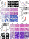
Overexpressed humanized circELP2/ miR‐630 mimic was sprayed into the mouse lung to evaluate circELP2 as a therapeutic target and circELP2‐mediated signal pathway. (A) qRT‐PCR result validated that overexpressed humanized circELP2 increased circELP2 in mice lung. (B) MicroCT imaging system for small animal clarified that the fibrosis degree was evidently higher in highly expressed circELP2 group than control group. (C) Overexpressed circELP2 treatment worsened lung function. (D) Images of H&E and Masson's trichrome staining depicted that the highly expressed circELP2 group had more collagen accumulation, more thickened alveolar walls and more deteriorate fibrotic degree than the control group. (E) Western blot unveiled that the fibrotic proteins were strengthened in highly expressed circELP2 group. The targeting proteins YAP1, TAZ, mTOR and mLST8 also increased in overexpressed circELP2‐treated mice. (F) qRT‐PCR result proved that miR‐630 increased in miR‐630 mimic‐treated mice. (G) The fibrotic proteins and the targeting proteins YAP1 and TAZ decreased in miR‐630 mimic‐treated mice. (H) miR‐630 mimic treatment promoted lung function. (I) MicroCT images demonstrated that the degree of fibrosis in miR‐630 mimic‐treated group was significantly lower than control group. (J) Immunohistochemical images depicted that miR‐630 mimic treatment improved lung structure and reduced collagen deposition.

Similar articles

Cited by

References

    1. Raghu G, Remy‐Jardin M, Richeldi L, et al. Idiopathic pulmonary fibrosis (an update) and progressive pulmonary fibrosis in adults: an official ATS/ERS/JRS/ALAT clinical practice guideline. Am J Respir Crit Care Med. 2022;205:e18‐e47. - PMC - PubMed
    1. Jia Y, Li X, Nan A, et al. Circular RNA 406961 interacts with ILF2 to regulate PM2.5‐induced inflammatory responses in human bronchial epithelial cells via activation of STAT3/JNK pathways. Environ Int. 2020;141:105755. - PubMed
    1. Li J, Li P, Zhang G, Qin P, Zhang D, Zhao W. CircRNA TADA2A relieves idiopathic pulmonary fibrosis by inhibiting proliferation and activation of fibroblasts. Cell Death Dis. 2020;11:553. - PMC - PubMed
    1. Zhou Z, Jiang R, Yang X, et al. circRNA mediates silica‐induced macrophage activation via HECTD1/ZC3H12A‐dependent ubiquitination. Theranostics. 2018;8:575‐592. - PMC - PubMed
    1. Chen X, Mao R, Su W, et al. Circular RNA circHIPK3 modulates autophagy via MIR124‐3p‐STAT3‐PRKAA/AMPKalpha signaling in STK11 mutant lung cancer. Autophagy. 2020;16:659‐671. - PMC - PubMed

Publication types

MeSH terms

Substances

LinkOut - more resources