

CTSB Nuclear Translocation Facilitates DNA Damage and Lysosomal Stress to Promote Retinoblastoma Cell Death
- PMID: 38159170
- PMCID: PMC11424708
- DOI: 10.1007/s12033-023-01042-0
CTSB Nuclear Translocation Facilitates DNA Damage and Lysosomal Stress to Promote Retinoblastoma Cell Death
Abstract
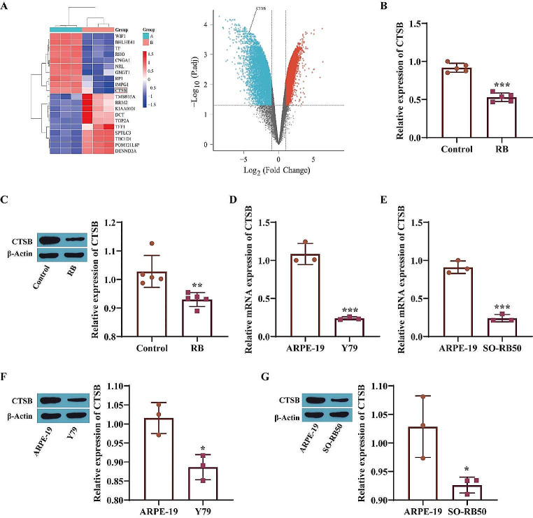
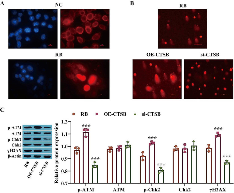
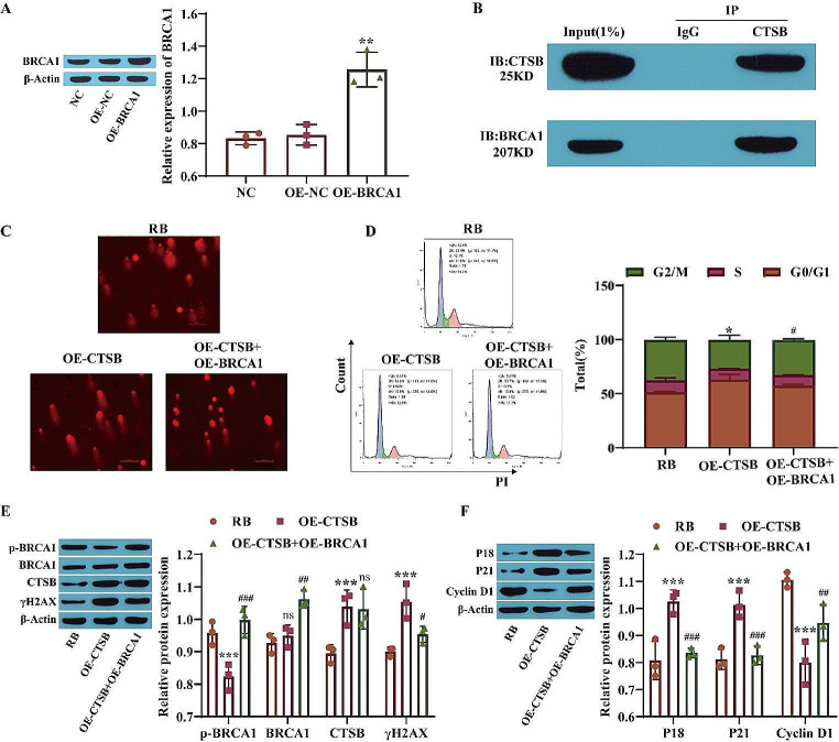
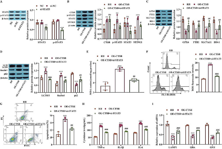
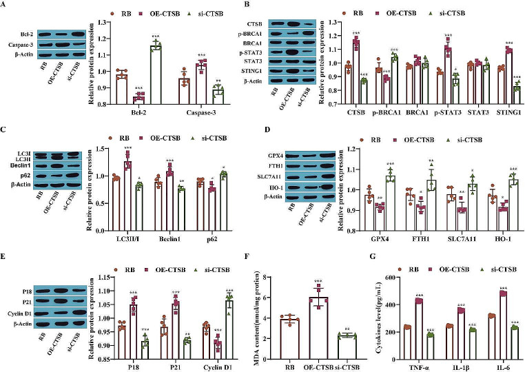
Retinoblastoma (RB) is a pernicious tumor originating from photoreceptor precursor cells that often endangers the lives of children. The purpose of our study was to further investigate the influence of cathepsin B (CTSB) nuclear translocation on RB cell death. Y79 cells were injected into the vitreous cavity of nude mice at a dose of 4 µL/mouse to establish an animal model of RB. Real-time quantitative polymerase chain reaction (RT-qPCR), Western blot analysis, a comet assay, a Cell Counting Kit-8 (CCK-8) assay and flow cytometry were used to measure the levels of the interrelated genes and proteins and to evaluate alterations in autophagy, apoptosis, proliferation, DNA damage and cell cycle arrest. CTSB was found to be expressed at low levels in RB animal model samples and RB cell lines. Functionally, CTSB nuclear translocation promoted DNA damage, cell cycle arrest, ferroptosis and autophagy in Y79 cells and inhibited their proliferation. Downstream mechanistic studies showed that nuclear translocation of CTSB facilitates DNA damage and cell cycle arrest in RB cells by inhibiting breast cancer 1 protein (BRCA1) expression and also activates the signal transducer and activator of transcription 3/stimulator of interferon response cGAMP interactor 1 (STAT3/STING1) pathway to induce lysosomal stress, leading to ferroptosis and autophagy in Y79 cells and alleviating RB. Nuclear translocation of CTSB facilitates DNA damage and cell cycle arrest in RB cells by inhibiting BRCA1 expression and activating the STAT3/STING1 pathway and induces lysosomal stress, which eventually leads to ferroptosis and autophagy and mitigates RB.
Keywords: Autophagy; CTSB nuclear translocation; DNA damage repair; Ferroptosis; Lysosomal stress; Retinoblastoma.
© 2023. The Author(s).
Conflict of interest statement
The authors declare that they have no competing financial interests.
Figures

Similar articles
-
Cathepsin B is a mediator of organelle-specific initiation of ferroptosis.Biochem Biophys Res Commun. 2020 Dec 17;533(4):1464-1469. doi: 10.1016/j.bbrc.2020.10.035. Epub 2020 Oct 22. Biochem Biophys Res Commun. 2020. PMID: 33268027
-
Targeting the lysosome by an aminomethylated Riccardin D triggers DNA damage through cathepsin B-mediated degradation of BRCA1.J Cell Mol Med. 2019 Mar;23(3):1798-1812. doi: 10.1111/jcmm.14077. Epub 2018 Dec 18. J Cell Mol Med. 2019. PMID: 30565390 Free PMC article.
-
MiR-452-5p facilitates retinoblastoma cell growth and invasion via the SOCS3/JAK2/STAT3 pathway.J Biochem Mol Toxicol. 2023 Dec;37(12):e23501. doi: 10.1002/jbt.23501. Epub 2023 Aug 26. J Biochem Mol Toxicol. 2023. PMID: 37632310
-
The role of STAT3 in autophagy.Autophagy. 2015;11(5):729-39. doi: 10.1080/15548627.2015.1017192. Autophagy. 2015. PMID: 25951043 Free PMC article. Review.
-
Cathepsin B induces kidney diseases through different types of programmed cell death.Front Immunol. 2025 Mar 10;16:1535313. doi: 10.3389/fimmu.2025.1535313. eCollection 2025. Front Immunol. 2025. PMID: 40129990 Free PMC article. Review.
Cited by
-
Airway specific deregulation of asthma-related serpins impairs tracheal architecture and oxygenation in D. melanogaster.Sci Rep. 2024 Jul 17;14(1):16567. doi: 10.1038/s41598-024-66752-0. Sci Rep. 2024. PMID: 39019933 Free PMC article.
-
Decoding ferroptosis: transforming orthopedic disease management.Front Pharmacol. 2024 Dec 6;15:1509172. doi: 10.3389/fphar.2024.1509172. eCollection 2024. Front Pharmacol. 2024. PMID: 39712490 Free PMC article. Review.
-
From mitochondrial dysregulation to ferroptosis: Exploring new strategies and challenges in radioimmunotherapy (Review).Int J Oncol. 2025 Sep;67(3):76. doi: 10.3892/ijo.2025.5781. Epub 2025 Aug 8. Int J Oncol. 2025. PMID: 40776761 Free PMC article. Review.
-
Antimicrobial peptide CRAMP/LL-37 mediates ferroptosis resistance in cardiomyocytes by inhibiting cathepsin L.Basic Res Cardiol. 2025 Jun 15. doi: 10.1007/s00395-025-01122-z. Online ahead of print. Basic Res Cardiol. 2025. PMID: 40517353
-
Ferroptosis: a novel mechanism of cell death in ophthalmic conditions.Front Immunol. 2024 Jun 27;15:1440309. doi: 10.3389/fimmu.2024.1440309. eCollection 2024. Front Immunol. 2024. PMID: 38994366 Free PMC article. Review.
References
MeSH terms
Substances
LinkOut - more resources
Full Text Sources
Research Materials
Miscellaneous
