. 2024 Jan 19;19(1):193-207.
doi: 10.1021/acschembio.3c00654. Epub 2023 Dec 30.

Identification of Protein Targets of S-Nitroso-Coenzyme A-Mediated S-Nitrosation Using Chemoproteomics

Affiliations

Identification of Protein Targets of S-Nitroso-Coenzyme A-Mediated S-Nitrosation Using Chemoproteomics

Julia A Falco et al. ACS Chem Biol. .

Abstract

S-Nitrosation is a cysteine post-translational modification fundamental to cellular signaling. This modification regulates protein function in numerous biological processes in the nervous, cardiovascular, and immune systems. Small molecule or protein nitrosothiols act as mediators of NO signaling by transferring the NO group (formally NO+) to a free thiol on a target protein through a transnitrosation reaction. The protein targets of specific transnitrosating agents and the extent and functional effects of S-nitrosation on these target proteins have been poorly characterized. S-nitroso-coenzyme A (CoA-SNO) was recently identified as a mediator of endogenous S-nitrosation. Here, we identified direct protein targets of CoA-SNO-mediated transnitrosation using a competitive chemical-proteomic approach that quantified the extent of modification on 789 cysteine residues in response to CoA-SNO. A subset of cysteines displayed high susceptibility to modification by CoA-SNO, including previously uncharacterized sites of S-nitrosation. We further validated and functionally characterized the functional effects of S-nitrosation on the protein targets phosphofructokinase (platelet type), ATP citrate synthase, and ornithine aminotransferase.


Figures

Figure 1.
Figure 1.
Identifying cysteine residues sensitive to CoA-SNO mediated S-nitrosation. (A) CoA-SNO is prepared by the reaction of CoA and sodium nitrite under acidic conditions. (B) Competitive cysteine-profiling strategy: Cell lysates are treated with a nitrosating agent (e.g. CoA-SNO) or a buffer control, labeled with the IA probe, and subjected to CuAAC to append either the Azo-L (CoA-SNO) or Azo-H (buffer). Labeled proteomes are mixed, subjected to streptavidin enrichment, on-bead trypsin digestion, and cleaved from beads and analyzed by high-resolution LC/LC-MS/MS to identify the (MS2) and quantify (MS1) the probe-labeled cysteines in each sample. (C) 789 cysteines identified and ranked by their sensitivity to CoA-SNO treatment (H:L ratio). Inset, 50 cysteines with the highest sensitivity to CoA-SNO mediated modification. (D) Representative extracted ion chromatograms and isotopic envelopes (light, red; heavy, blue) are shown for three cysteines with low, moderate, and high sensitivity to CoA-SNO.
Figure 2.
Figure 2.
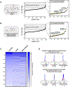
Assessing the selectivity of nitrosation donors: GSNO, PAPA NONOate, and CoA-SNO. (A) 1038 cysteines identified and ranked according to the extent of modification by the transnitrosating agent GSNO. Inset shows the 50 most sensitive cysteines to GSNO treatment. (B) 985 cysteines identified and ranked according to the extent of modification by the NO donor, PAPA NONOate. Inset shows 50 most sensitive cysteines to PAPA NONOate treatment. (C) Heatmap comparing the sensitivity of 489 individual cysteine residues to each of the NO donors: CoA-SNO, GSNO, and PAPA NONOate. Darker blue indicates higher sensitivity to the corresponding NO donor. (D) Representative extracted ion chromatograms display cysteines with differential sensitivity to the various NO donors, with Cys329 of CTSD being highly sensitive to CoA-SNO, GSNO, and PAPA NONOate and Cys845 of ACLY being highly sensitive to CoA-SNO, but insensitive to GSNO or PAPA NONOate.
Figure 3.
Figure 3.
Functional characterization of S-nitrosation at Cys360 in PFKP. (A) PFKP catalyzes the conversion of fructose-6-phosphate (F-6-P) to fructose-1,6-phosphate (F-1,6-P) using ATP. (B) Treatment of PFKP WT with increasing concentrations of CoA-SNO (0–30 μM) shows increased levels of nitrosated PFKP monitored by the biotin switch assay. (C) Nitrosation of PFKP by 30 μM CoA-SNO is dependent on Cys360 with undetectable S-nitrosation in the PFKP cysteine 360 to serine mutant (C360S) monitored by the biotin switch assay. (D) Treatment of PFKP with 30 μM CoA-SNO, GSNO, and MAHMA NONOate results in nitrosated PFKP formation as monitored by the biotin switch assay. (E) Structure of PFKP in its active tetrameric form with Cys360 located near the ATP binding site (inset) (PDB ID: 4RH3). (F) Nitrosation of PFKP inhibits enzymatic activity in the WT protein while the mutant C360S is resistant to activity changes (* = significant, p < 0.05; ns = not significant).
Figure 4.
Figure 4.
Functional characterization of S-nitrosation at Cys845 in ACLY. (A) ACLY catalyzes the reaction of citrate and CoA to oxaloacetate and acetyl-CoA using ATP. (B) Treatment of ACLY WT with increasing concentrations of CoA-SNO (0–30 μM) shows increased levels of nitrosated ACLY monitored by the biotin switch assay. (C) Nitrosation of ACLY by 30 μM CoA-SNO depends on Cys845 with undetectable S-nitrosation in the ACLY cysteine 845 to serine mutant (C845S) monitored by the biotin switch assay. (D) Treatment of ACLY with 30 μM CoA-SNO, GSNO, and MAHMA NONOate results in nitrosated ACLY formation as monitored by the biotin switch assay. (E) Homotetrameric structure of ACLY (PDB ID: 6O0H). Cys845 is located within the citrate binding domain at the dimer interface. (F) Nitrosation of ACLY inhibits enzymatic activity in the WT protein, while the mutant C845S is more resistant to activity changes. (G) Inhibition of ACLY WT and C845S by increasing concentrations of GSNO (0–500 μM).
Figure 5.
Figure 5.
Functional characterization of S-nitrosation at Cys150 in OAT. (A) OAT catalyzes the transformation of α-ketoglutarate and ornithine to glutamate and glutamate-5-semialdehyde. (B) Treatment of OAT WT with increasing concentrations of CoA-SNO (0–30 μM) shows increased levels of nitrosated OAT monitored by the biotin switch assay. (C) Nitrosation of OAT by 30 μM CoA-SNO is dependent on Cys150 with undetectable S-nitrosation in the OAT cysteine 150 to serine mutant (C150S) monitored by the biotin switch assay. (D) Treatment of OAT with 30 μM CoA-SNO, GSNO, and MAHMA NONOate results in nitrosated OAT formation as monitored by the biotin switch assay. (E) OAT exists as a homodimer with Cys150 located near the PLP cofactor binding site (PDB ID: 1OAT). (F) Nitrosation of OAT inhibits enzymatic activity in the WT and C150A protein (ns = not significant, p > 0.05).

Similar articles

References

    1. Martínez MC; Andriantsitohaina R Reactive Nitrogen Species: Molecular Mechanisms and Potential Significance in Health and Disease. Antioxidants and Redox Signaling. Mary Ann Liebert Inc. March 1, 2009, pp 669–702. 10.1089/ars.2007.1993. - DOI - PubMed
    1. Villanueva C; Giulivi C Subcellular and Cellular Locations of Nitric Oxide Synthase Isoforms as Determinants of Health and Disease. Free Radical Biology and Medicine. Pergamon: August 1, 2010, pp 307–316. 10.1016/j.freeradbiomed.2010.04.004. - DOI - PMC - PubMed
    1. Shahani N; Sawa A Protein S-Nitrosylation: Role for Nitric Oxide Signaling in Neuronal Death. Biochimica et Biophysica Acta - General Subjects. Elsevier; June 1, 2012, pp 736–742. 10.1016/j.bbagen.2011.07.010. - DOI - PMC - PubMed
    1. Calabrese V; Cornelius C; Rizzarelli E; Owen JB; Dinkova-Kostova AT; Butterfield DA Nitric Oxide in Cell Survival: A Janus Molecule. Antioxidants and Redox Signaling. Mary Ann Liebert, Inc. 140 Huguenot Street, 3rd Floor New Rochelle, NY 10801 USA November 1, 2009, pp 2717–2739. 10.1089/ars.2009.2721. - DOI - PubMed
    1. Knott AB; Bossy-Wetzel E Nitric Oxide in Health and Disease of the Nervous System. Antioxidants and Redox Signaling. Mary Ann Liebert, Inc. 140 Huguenot Street, 3rd Floor New Rochelle, NY 10801 USA March 1, 2009, pp 541–553. 10.1089/ars.2008.2234. - DOI - PMC - PubMed

Publication types

LinkOut - more resources