. 2024 Feb;300(2):105608.
doi: 10.1016/j.jbc.2023.105608. Epub 2023 Dec 28.

Probing the mechanism by which the retinal G protein transducin activates its biological effector PDE6

Affiliations

Probing the mechanism by which the retinal G protein transducin activates its biological effector PDE6

Cody Aplin et al. J Biol Chem. 2024 Feb.

Abstract

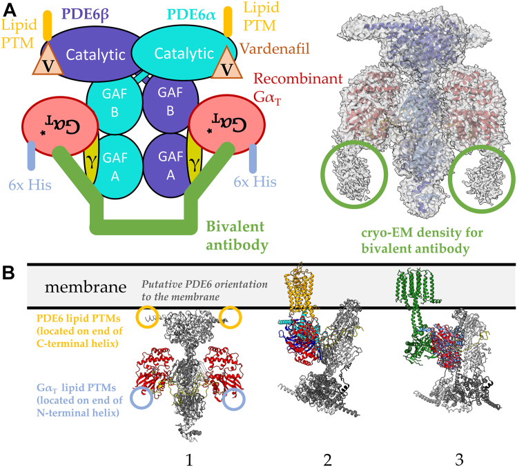
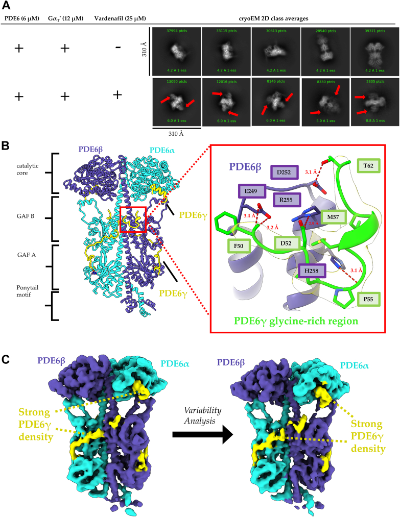
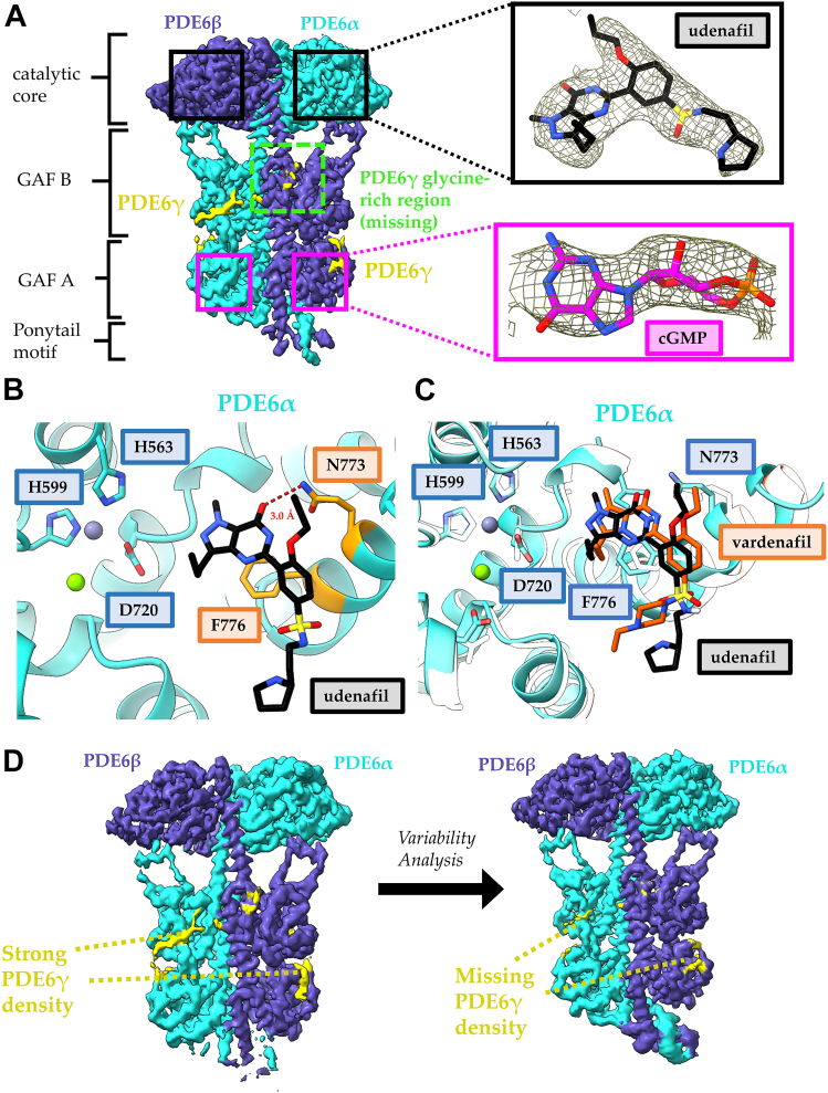
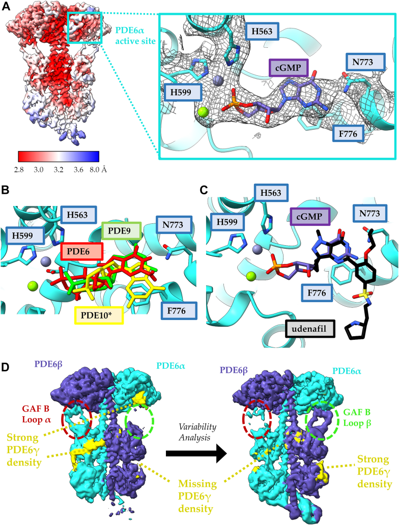
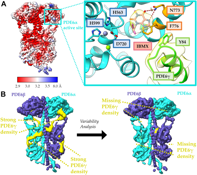
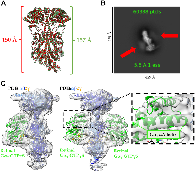
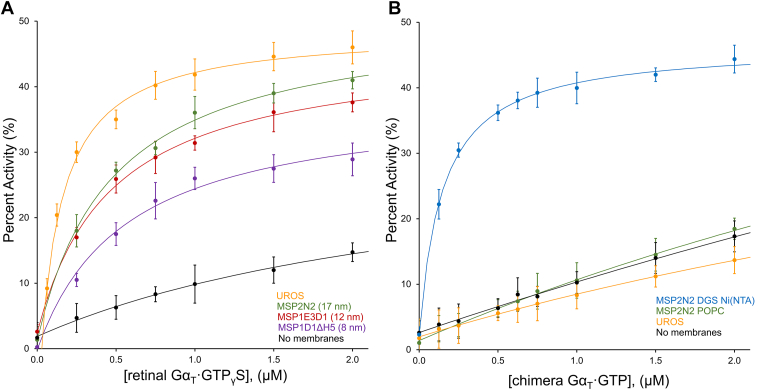
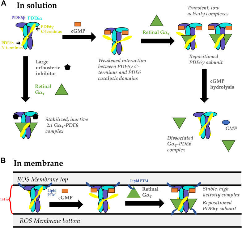
Phototransduction in retinal rods occurs when the G protein-coupled photoreceptor rhodopsin triggers the activation of phosphodiesterase 6 (PDE6) by GTP-bound alpha subunits of the G protein transducin (GαT). Recently, we presented a cryo-EM structure for a complex between two GTP-bound recombinant GαT subunits and native PDE6, that included a bivalent antibody bound to the C-terminal ends of GαT and the inhibitor vardenafil occupying the active sites on the PDEα and PDEβ subunits. We proposed GαT-activated PDE6 by inducing a striking reorientation of the PDEγ subunits away from the catalytic sites. However, questions remained including whether in the absence of the antibody GαT binds to PDE6 in a similar manner as observed when the antibody is present, does GαT activate PDE6 by enabling the substrate cGMP to access the catalytic sites, and how does the lipid membrane enhance PDE6 activation? Here, we demonstrate that 2:1 GαT-PDE6 complexes form with either recombinant or retinal GαT in the absence of the GαT antibody. We show that GαT binding is not necessary for cGMP nor competitive inhibitors to access the active sites; instead, occupancy of the substrate binding sites enables GαT to bind and reposition the PDE6γ subunits to promote catalytic activity. Moreover, we demonstrate by reconstituting GαT-stimulated PDE6 activity in lipid bilayer nanodiscs that the membrane-induced enhancement results from an increase in the apparent binding affinity of GαT for PDE6. These findings provide new insights into how the retinal G protein stimulates rapid catalytic turnover by PDE6 required for dim light vision.

Keywords: G protein; cryoelectron microscopy; phosphodiesterase; phototransduction; signal transduction; structural biology.


Conflict of interest statement

Conflict of interest The authors declare that they have no conflict of interest with the contents of this article.

Figures

Figure 1
Figure 1
Depictions of the interactions of GTP-bound transducin Gα subunits with PDE6.A, left: the cryo-EM structure of the GαT∗–PDE6 complex solved by Gao et al. was engineered using recombinant GαT∗ subunits, a bivalent antibody, and the orthosteric inhibitor vardenafil (PDB: 7JSN). Right: weak cryo-EM density for the bivalent antibody is observed in the cryo-EM map of GαT∗–PDE6 complex (red circles, EMDB: 22,458). B, comparison of the orientation of the Gα subunit in different protein complexes. 1, the presumed association of the 2:1 GαT–PDE6 complex with the membrane, based on the location of lipid modifications on the C-terminal helices of the PDE6α and PDE6β subunits (yellow). The upside-down orientation of GαT∗ is highlighted by labeling the location of the N-terminal helix of GαT∗, which contains PTMs in native retinal GαT (blue). 2, orientation of the GαT subunit in the cryo-EM structure of the rhodopsin–transducin complex (PDB: 6OY9) compared to the GαT–PDE6 complex. One GαT subunit is shown in the GαT–PDE6 complex for simplicity. The structures are aligned by the GαT subunits and membrane-bound rhodopsin is used to orient the complexes with the membrane. 3, orientation of the Gαs subunit in the cryo-EM structure of adenylyl cyclase bound to Gαs (PDB: 6R3Q) compared to the GαT–PDE6 complex. One GαT subunit is shown in the GαT–PDE6 complex for simplicity. The structures are aligned by the Gα subunits and membrane-bound adenylyl cyclase is used to orient the complexes with the membrane. PDE, phosphodiesterase; PTM, posttranslational lipid modification.
Figure 2
Figure 2
The glycine-rich region of PDE6γ binds near the PDE6 dimerization interface.A, comparison of 2D classes of the GαT∗–PDE6 complex, with and without vardenafil treatment. In the presence of vardenafil, the GαT∗–PDE6 complex forms at a 2:1 stoichiometric ratio and does not readily form in its absence. The GαT∗ subunit is indicated by the red arrows. B, the improved cryo-EM structure of the PDE6 heterotetramer. The PDE6α (cyan), PDE6β (purple), and PDE6γ (yellow) are denoted and the domains of the PDE6 holoenzyme are labeled. The hydrogen bonding network between the PDE6γ glycine-rich region (green) and PDE6β (purple) is shown in the insert (red) with the cryo-EM density (yellow). C, 3DVA shows that PDE6γ remains tightly associated with the PDE6α and PDE6β across all variability components (also see Fig. S3). 3DVA, 3D variability analysis; PDE, phosphodiesterase.
Figure 3
Figure 3
Bulky, orthosteric PDE inhibitors disrupt PDE6γ binding by sterically competing with the PDE6γ C terminus.A, 2.9 Å cryo-EM map of PDE6–udenafil complex. The map density for the PDE6α (cyan), PDE6β (purple), and PDE6γ (yellow) are colored within 4 Å of the atomic model and the location of the udenafil and cGMP binding sites are highlighted with black and magenta boxes, respectively. Inset: strong cryo-EM map density is shown for udenafil (black) bound at the active site and cGMP (magenta) bound to the GAF A domain. The missing density for the PDE6γ glycine-rich region is labeled in a dashed box (green). B, the udenafil (black) binding pocket in the catalytic core of PDE6α. Magnesium (green) and zinc (gray) are shown as spheres in the active site and the residues important for metal ion coordination are labeled. The strongest interactions between udenafil and PDE6 are π–π stacking with F776 and hydrogen bonding (red) with N773 which are highlighted in orange. C, comparison of the ligand poses for vardenafil (PDB: 7JSN, orange) and udenafil (this paper, black) in the PDE6α active site. PDE6α from the udenafil-bound structure is colored cyan and PDE6α from the vardenafil-bound structure is colored white. Magnesium (green) and zinc (gray) are shown as spheres in the active site and the residues important for metal ion coordination are labeled. D, 3DVA analysis of the PDE6–udenafil complex shows a diminished association of PDE6γ with the GAF domains PDE6αβ across all variability components. The map density for the PDE6α (cyan), PDE6β (purple), and PDE6γ (yellow) are colored within 5 Å of the atomic model. 3DVA, 3D variability analysis; cGMP, cyclic GMP; PDE, phosphodiesterase.
Figure 4
Figure 4
cGMP binding disrupts PDE6γ binding without a steric clash and without the need for GαT.A, local resolution map of the PDE6–cGMP complex, displaying resolution estimates from 2.8 to 8 Å. The inset shows cryo-EM map density for the active site of PDE6α (cyan) when cGMP is bound. The density is displayed within 3 Å of the labeled residues. Magnesium (green) and zinc (gray) are shown as spheres in the active site and the residues important for metal ion coordination are labeled. B, comparison of the docking of cGMP into PDE6 (red), an inhibited PDE9 (green), and a catalytically dead PDE10 mutant (yellow). The ligand poses for cGMP in PDE6 and PDE9 are very similar; however, the PDE10 mutation exhibits a different docking pose. C, comparison of the ligand poses of cGMP and udenafil in the PDE6α (cyan) active site. The heterocyclic rings of both ligands are positioned in the same manner for π–π stacking with F776 and form hydrogen bonds with N773. D, 3DVA analysis of the PDE6–cGMP complex shows a diminished association between PDE6γ and the PDE6α and PDE6β catalytic subunits across all variability components. The map density for the PDE6α (cyan), PDE6β (purple), and PDE6γ (yellow) are colored within 5 Å of the atomic model. 3DVA, 3D variability analysis; cGMP, cyclic GMP; PDE, phosphodiesteras.
Figure 5
Figure 5
A small orthosteric inhibitor, IBMX, disrupts PDE6γ binding without a steric clash with the PDE6γ C terminus.A, local resolution map of the PDE6–IBMX complex, displaying resolution estimates from 2.9 to 8 Å. The inset shows the IBMX-binding site in the PDE6–IBMX complex. IBMX is stabilized by π–π stacking with F776 and hydrogen bonding (red) with N773, which are highlighted in orange. Strong cryo-EM density for the PDE6γ subunit and IBMX is shown in yellow. Magnesium (green) and zinc (gray) are shown as spheres in the active site and the residues important for metal ion coordination are labeled. B, 3DVA analysis of the PDE6–IBMX complex shows a diminished association of PDE6γ with the GAF domains PDE6αβ across all variability components. The map density for the PDE6α (cyan), PDE6β (purple), and PDE6γ (yellow) are colored within 5 Å of the atomic model. 3DVA, 3D variability analysis; IBMX, inhibitor 3-isobutyl 1-methylxanthine; PDE, phosphodiesterase.
Figure 6
Figure 6
Retinal GαT forms a PDE6-GαT complex with both 1:1 and 1:2 stoichiometric ratios in the presence of IBMX.A, IBMX-bound PDE6 (green) is elongated compared to apo-PDE6 (red). The distance was measured between residues Glu8 and Glu510 of the PDE6β subunit. B, 2D class of IBMX-bound PDE6 in the presence of retinal GαT·GTPγS. Complex formation is diminished in comparison to vardenafil treatment; however, weak density for GαT is observed (red arrows). C, low-resolution cryo-EM maps of the retinal GαT·GTPγS–PDE6 complex with both 1:1 and 1:2 stoichiometries. The cryo-EM map density for GαT·GTPγS is strong for the αA helix of GαT allowing for the determination of its upside-down orientation relative to PDE6 (see Figs. 1B and S5). IBMX, inhibitor 3-isobutyl 1-methylxanthine; PDE, phosphodiesterase.
Figure 7
Figure 7
Transducin-stimulated PDE6 activity in the presence of lipid nanodiscs.A, retinal GαT·GTPγS–stimulated PDE6 activity in the presence of UROS (orange), MSP2N2 nanodiscs (green), MSP1E3D1 nanodiscs (red), and MSP1D1Δh5 nanodiscs (purple). A control without membranes is included for comparison (black). All nanodiscs contain 100% POPC lipids. Data is shown as the average of three independent experiments with the SD shown as error bars (n = 3). B, chimera GαT∗-stimulated PDE6 activity in the presence of UROS (orange), MSP2N2 POPC nanodiscs (100% POPC, green), and MSP2N2 POPC DGS Ni(NTA) nanodiscs (95% POPC, 5% DGS Ni(NTA), blue). A control without membranes is included for comparison (black). Data is shown as the average of three independent experiments with the SD shown as error bars (n = 3). PDE, phosphodiesterase; POPC, 1-palmitoyl-2-oleoyl-glycero-3-phosphocholine.
Figure 8
Figure 8
Activation mechanism of PDE6 by GαT.A, in a solution environment, cGMP is able to bind at the PDE6 catalytic sites, loosening the binding of PDE6γ. This promotes the recruitment of GαT by the PDE6γ C terminus, resulting in the formation of transient, low activity GαT–PDE6 complexes with both 1:1 and 2:1 stoichiometric ratios. These complexes dissociate after hydrolysis. In the presence of large orthosteric inhibitors, such as vardenafil, an inactive but stable 2:1 GαT–PDE6 complex forms. B, in a membrane environment, retinal GαT subunits use lipid PTM to anchor to the adjacent membrane promoting the formation of high-affinity, stable GαT–PDE6 complexes with displaced PDE6γ subunits, driving maximal PDE6 catalytic activity. cGMP, cyclic GMP; PDE, phosphodiesterase; PTM, posttranslational lipid modification.

Similar articles

Cited by

References

    1. Burns M.E., Arshavsky V.Y. Beyond counting photons: trials and trends in vertebrate visual transduction. Neuron. 2005;48:387–401. - PubMed
    1. Palczewski K. G protein-coupled receptor rhodopsin. Annu. Rev. Biochem. 2006;75:743–767. - PMC - PubMed
    1. Hayashi F., Saito N., Tanimoto Y., Okada K., Morigaki K., Seno K., et al. Raftophilic rhodopsin-clusters offer stochastic platforms for G protein signalling in retinal discs. Commun. Biol. 2019;2:209. - PMC - PubMed
    1. Baillie G.S., Tejeda G.S., Kelly M.P. Therapeutic targeting of 3′,5′-cyclic nucleotide phosphodiesterases: inhibition and beyond. Nat. Rev. Drug Discov. 2019;18:770–796. - PMC - PubMed
    1. Maurice D.H., Ke H., Ahmad F., Wang Y., Chung J., Manganiello V.C. Advances in targeting cyclic nucleotide phosphodiesterases. Nat. Rev. Drug Discov. 2014;13:290–314. - PMC - PubMed

Publication types

MeSH terms

LinkOut - more resources