

Probing the mechanism by which the retinal G protein transducin activates its biological effector PDE6
- PMID: 38159849
- PMCID: PMC10838916
- DOI: 10.1016/j.jbc.2023.105608
Probing the mechanism by which the retinal G protein transducin activates its biological effector PDE6
Abstract
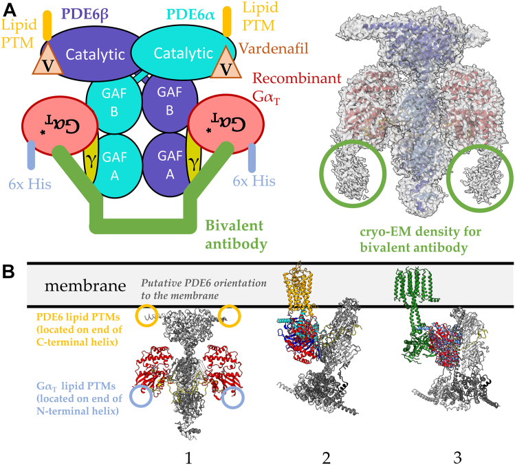
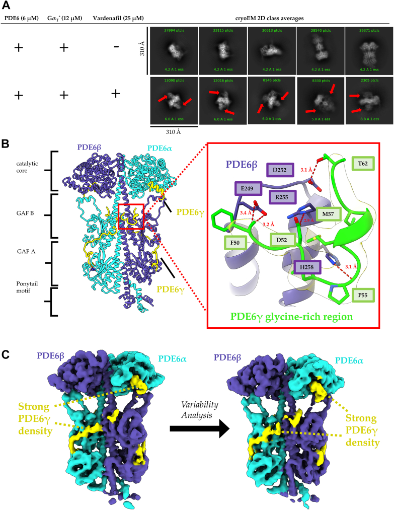
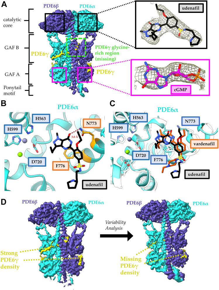
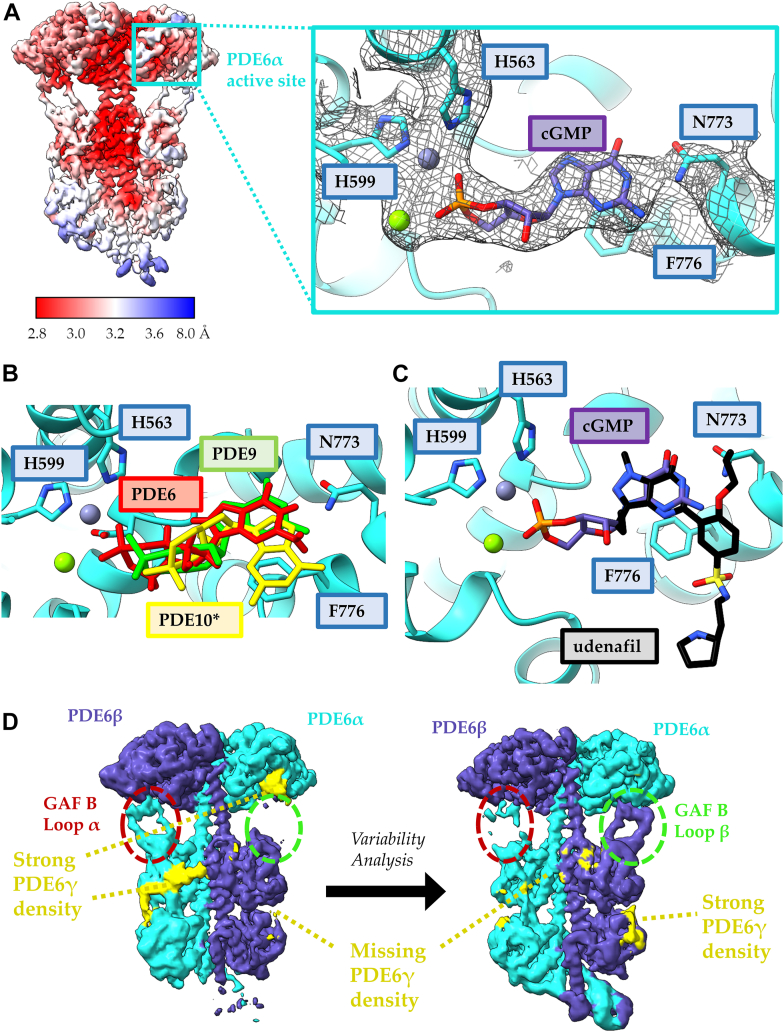
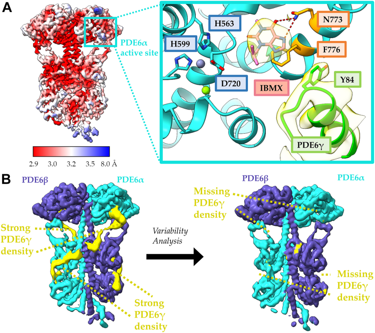
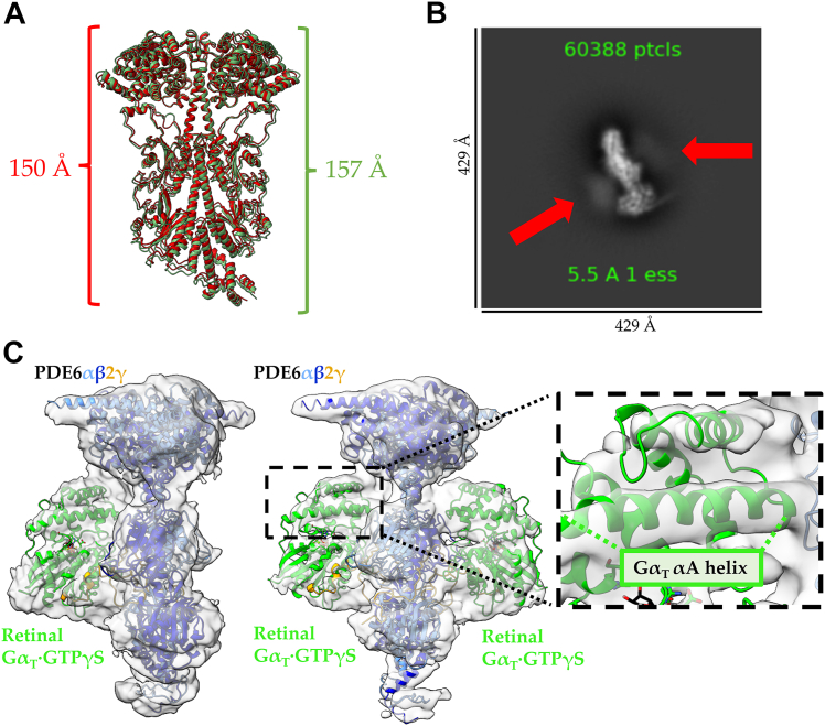
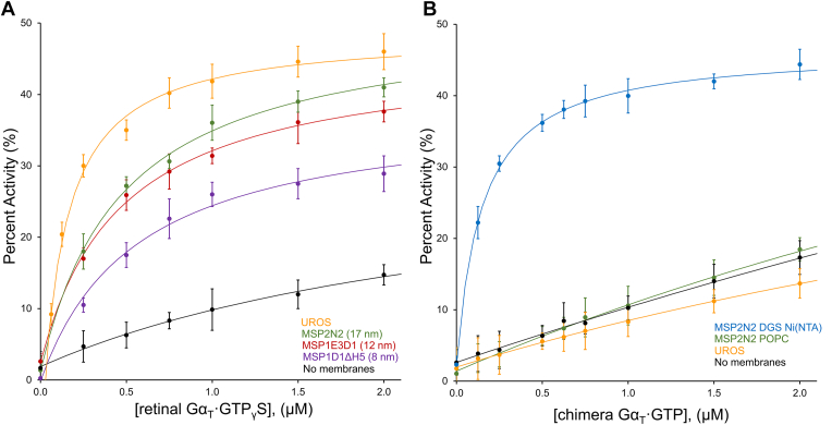
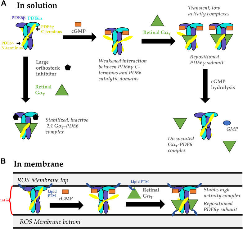
Phototransduction in retinal rods occurs when the G protein-coupled photoreceptor rhodopsin triggers the activation of phosphodiesterase 6 (PDE6) by GTP-bound alpha subunits of the G protein transducin (GαT). Recently, we presented a cryo-EM structure for a complex between two GTP-bound recombinant GαT subunits and native PDE6, that included a bivalent antibody bound to the C-terminal ends of GαT and the inhibitor vardenafil occupying the active sites on the PDEα and PDEβ subunits. We proposed GαT-activated PDE6 by inducing a striking reorientation of the PDEγ subunits away from the catalytic sites. However, questions remained including whether in the absence of the antibody GαT binds to PDE6 in a similar manner as observed when the antibody is present, does GαT activate PDE6 by enabling the substrate cGMP to access the catalytic sites, and how does the lipid membrane enhance PDE6 activation? Here, we demonstrate that 2:1 GαT-PDE6 complexes form with either recombinant or retinal GαT in the absence of the GαT antibody. We show that GαT binding is not necessary for cGMP nor competitive inhibitors to access the active sites; instead, occupancy of the substrate binding sites enables GαT to bind and reposition the PDE6γ subunits to promote catalytic activity. Moreover, we demonstrate by reconstituting GαT-stimulated PDE6 activity in lipid bilayer nanodiscs that the membrane-induced enhancement results from an increase in the apparent binding affinity of GαT for PDE6. These findings provide new insights into how the retinal G protein stimulates rapid catalytic turnover by PDE6 required for dim light vision.
Keywords: G protein; cryoelectron microscopy; phosphodiesterase; phototransduction; signal transduction; structural biology.
Copyright © 2024 The Authors. Published by Elsevier Inc. All rights reserved.
Conflict of interest statement
Conflict of interest The authors declare that they have no conflict of interest with the contents of this article.
Figures
References
-
- Burns M.E., Arshavsky V.Y. Beyond counting photons: trials and trends in vertebrate visual transduction. Neuron. 2005;48:387–401. - PubMed
Publication types
MeSH terms
Substances
Grants and funding
LinkOut - more resources
Full Text Sources
