An eNAMPT-neutralizing mAb reduces post-infarct myocardial fibrosis and left ventricular dysfunction
- PMID: 38160623
- PMCID: PMC10872269
- DOI: 10.1016/j.biopha.2023.116103
An eNAMPT-neutralizing mAb reduces post-infarct myocardial fibrosis and left ventricular dysfunction
Abstract
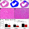
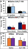
Myocardial infarction (MI) triggers adverse ventricular remodeling (VR), cardiac fibrosis, and subsequent heart failure. Extracellular nicotinamide phosphoribosyltransferase (eNAMPT) is postulated to play a significant role in VR processing via activation of the TLR4 inflammatory pathway. We hypothesized that an eNAMPT specific monoclonal antibody (mAb) could target and neutralize overexpressed eNAMPT post-MI and attenuate chronic cardiac inflammation and fibrosis. We investigated humanized ALT-100 and ALT-300 mAb with high eNAMPT-neutralizing capacity in an infarct rat model to test our hypothesis. ALT-300 was 99mTc-labeled to generate 99mTc-ALT-300 for imaging myocardial eNAMPT expression at 2 hours, 1 week, and 4 weeks post-IRI. The eNAMPT-neutralizing ALT-100 mAb (0.4 mg/kg) or saline was administered intraperitoneally at 1 hour and 24 hours post-reperfusion and twice a week for 4 weeks. Cardiac function changes were determined by echocardiography at 3 days and 4 weeks post-IRI. 99mTc-ALT-300 uptake was initially localized to the ischemic area at risk (IAR) of the left ventricle (LV) and subsequently extended to adjacent non-ischemic areas 2 hours to 4 weeks post-IRI. Radioactive uptake (%ID/g) of 99mTc-ALT-300 in the IAR increased from 1 week to 4 weeks (0.54 ± 0.16 vs. 0.78 ± 0.13, P < 0.01). Rats receiving ALT-100 mAb exhibited significantly improved myocardial histopathology and cardiac function at 4 weeks, with a significant reduction in the collagen volume fraction (%LV) compared to controls (21.5 ± 6.1% vs. 29.5 ± 9.9%, P < 0.05). Neutralization of the eNAMPT/TLR4 inflammatory cascade is a promising therapeutic strategy for MI by reducing chronic inflammation, fibrosis, and preserving cardiac function.
Keywords: Cardiac fibrosis; Cardioprotective effect; DAMP; Myocardial infarction; eNAMPT; mAb.
Copyright © 2023 The Authors. Published by Elsevier Masson SAS.. All rights reserved.
Conflict of interest statement
Declaration of Competing Interest Dr. Joe G.N. Garcia is CEO and Founder of Aqualung Therapeutics Corporation. All other authors report no conflict of interest or relationships relevant to the contents of this paper to disclose.
Figures


Similar articles
-
Extracellular Nicotinamide Phosphoribosyltransferase Is a Therapeutic Target in Experimental Necrotizing Enterocolitis.Biomedicines. 2024 Apr 28;12(5):970. doi: 10.3390/biomedicines12050970. Biomedicines. 2024. PMID: 38790933 Free PMC article.
-
eNAMPT Is a Novel Damage-associated Molecular Pattern Protein That Contributes to the Severity of Radiation-induced Lung Fibrosis.Am J Respir Cell Mol Biol. 2022 May;66(5):497-509. doi: 10.1165/rcmb.2021-0357OC. Am J Respir Cell Mol Biol. 2022. PMID: 35167418 Free PMC article.
-
Involvement of eNAMPT/TLR4 inflammatory signaling in progression of non-alcoholic fatty liver disease, steatohepatitis, and fibrosis.FASEB J. 2023 Mar;37(3):e22825. doi: 10.1096/fj.202201972RR. FASEB J. 2023. PMID: 36809677 Free PMC article.
-
Targeting the glucagon receptor improves cardiac function and enhances insulin sensitivity following a myocardial infarction.Cardiovasc Diabetol. 2019 Jan 9;18(1):1. doi: 10.1186/s12933-019-0806-4. Cardiovasc Diabetol. 2019. PMID: 30626440 Free PMC article.
-
Clinical aspects of left ventricular diastolic function assessed by Doppler echocardiography following acute myocardial infarction.Dan Med Bull. 2001 Nov;48(4):199-210. Dan Med Bull. 2001. PMID: 11767125 Review.
Cited by
-
Safety, Tolerability and Pharmacokinetics of the eNAMPT-Neutralizing ALT-100 Mab in Healthy Volunteers.J Clin Res Clin Trials. 2024 Sep;3(3):https://bioresscientia.com/article/safety-tolerability-and-pharmacokinetics-of-the-enampt-neutralizing-alt-100-mab-in-healthy-volunteers. Epub 2024 Sep 18. J Clin Res Clin Trials. 2024. PMID: 39950187 Free PMC article.
-
eNAMPT is a novel therapeutic target for mitigation of coronary microvascular disease in type 2 diabetes.Diabetologia. 2024 Sep;67(9):1998-2011. doi: 10.1007/s00125-024-06201-9. Epub 2024 Jun 19. Diabetologia. 2024. PMID: 38898303 Free PMC article.
-
TLR4 Ligation by eNAMPT, a Novel DAMP, is Essential to Sulfur Mustard- Induced Inflammatory Lung Injury and Fibrosis.Eur J Respir Med. 2024 Feb;6(1):389-397. Epub 2024 Jan 8. Eur J Respir Med. 2024. PMID: 38390523 Free PMC article.
-
Exploring the mechanism of rosmarinic acid in the treatment of lung adenocarcinoma based on bioinformatics methods and experimental validation.Discov Oncol. 2025 Jan 15;16(1):47. doi: 10.1007/s12672-025-01784-0. Discov Oncol. 2025. PMID: 39812944 Free PMC article.
-
NEDD4 E3 ligase-catalyzed NAMPT ubiquitination and autophagy activation are essential for pyroptosis-independent NAMPT secretion in human monocytes.Cell Commun Signal. 2025 Mar 30;23(1):157. doi: 10.1186/s12964-025-02164-5. Cell Commun Signal. 2025. PMID: 40159488 Free PMC article.
References
-
- Valikeserlis I, Athanasiou AA, Stakos D, Cellular mechanisms and pathways in myocardial reperfusion injury, Coron. Artery Dis. 32 (2021) 567–577. - PubMed
-
- Zhao ZQ, Velez DA, Wang NP, Hewan-Lowe KO, Nakamura M, Guyton RA, et al., Progressively developed myocardial apoptotic cell death during late phase of reperfusion, Apoptosis 6 (2001) 279–290. - PubMed
MeSH terms
Substances
Grants and funding
LinkOut - more resources
Full Text Sources
Medical

