

Elastofibroma Dorsi Detected Incidentally on Chest Computed Tomography: The Prevalence and Reporting Rate in Radiology Reports
- PMID: 38161569
- PMCID: PMC10757754
- DOI: 10.7759/cureus.51280
Elastofibroma Dorsi Detected Incidentally on Chest Computed Tomography: The Prevalence and Reporting Rate in Radiology Reports
Abstract
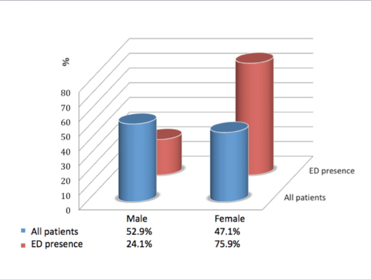
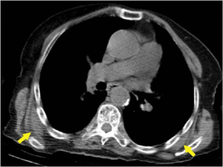
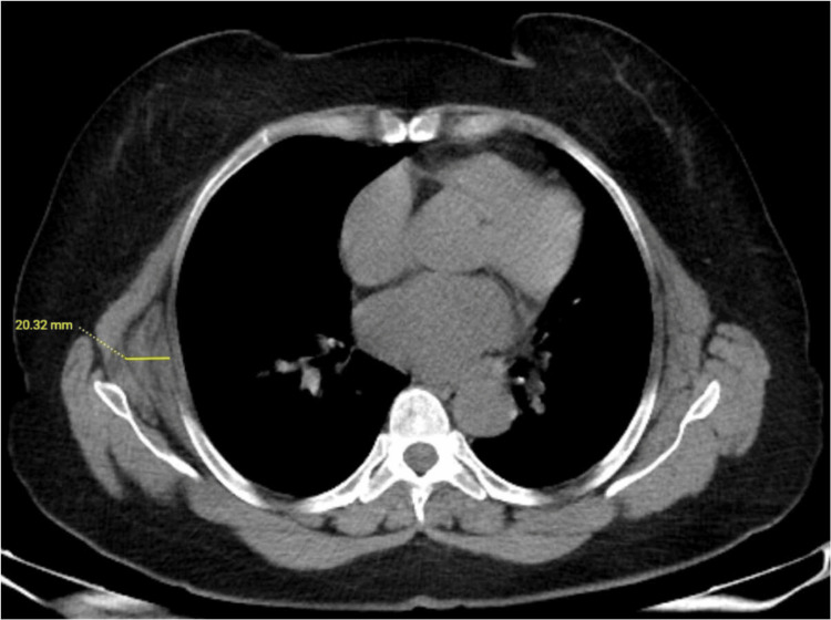
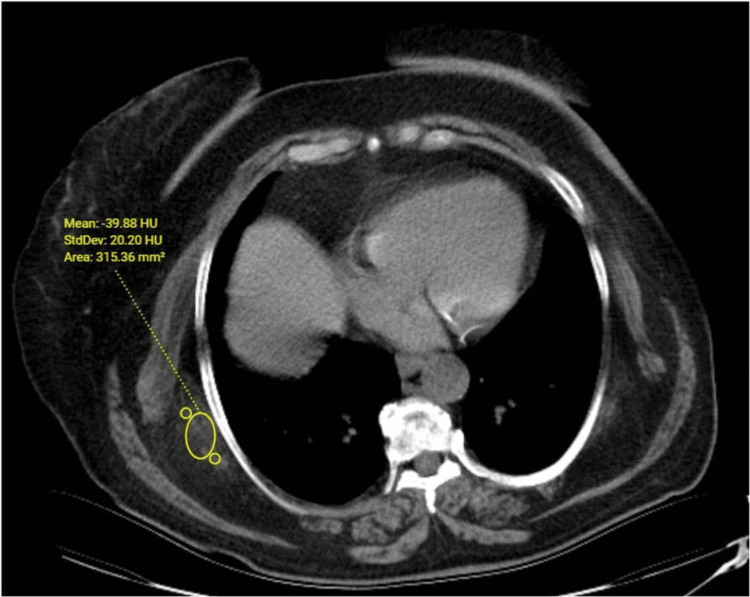
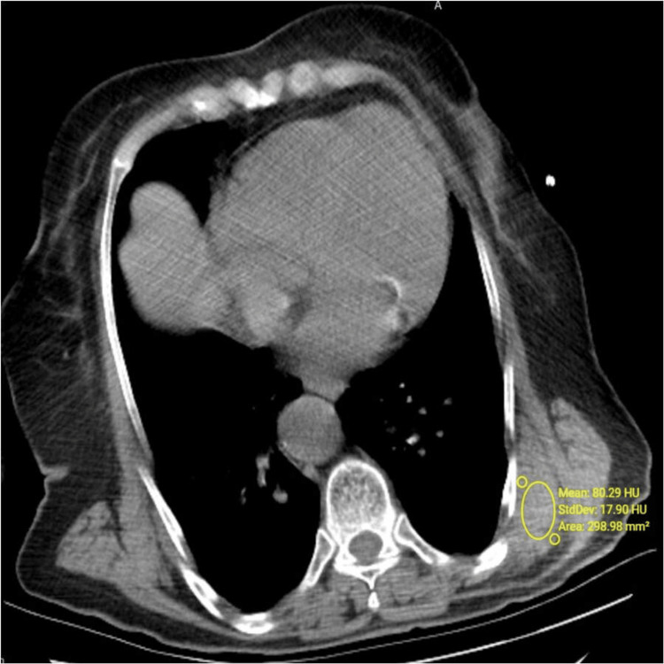
Background Elastofibroma dorsi (ED) is an uncommon benign connective-tissue tumor, usually seen in the subscapular region of women after the fifth decade. The study aimed to determine the prevalence, radiological characteristics, and the rates of mention in the initial radiology reports of ED incidentally detected by chest computed tomography (CT) imaging in a large population. Methodology This study was conducted at Hitit University, Erol Olçok Training and Research Hospital Radiology Department in Çorum, Turkey. A total of 3,299 patients (1,554 females, 1,745 males) who underwent non-contrast chest CT for various reasons were included in this retrospective study. The presence, side, thickness, and density of ED were investigated in these patients. Differences in gender and laterality were assessed statistically. It was also reviewed whether it was stated in the initial radiology report or not. Results ED was detected in 79 (2.4%) of 3,299 patients, in 60 (75.9%) females and 19 (24.1%) males with a median age of 57.5 years (range, 18-99 years). The risk of ED presence was determined to be 3.65-fold in females than in males. In the cases determined with ED, the median age was 75 years (range, 53-96 years), and ED was not determined in any patient aged <50 years. A statistically significant correlation was determined between age and the presence of ED (p < 0.001). No statistically significant correlation was found between age and ED thickness or density (p = 0.602, p = 0.233, respectively). It was noted that none of the patients were diagnosed in the first radiological report. Conclusions ED can be easily overlooked on cross-sectional examinations because of the similar appearance and density to adjacent structures. Knowledge of the characteristic radiological features of these lesions and increased awareness of radiologists will make early diagnosis possible in asymptomatic cases.
Keywords: computed tomography; density; elastofibroma dorsi; prevelance; radiology reports; thickness.
Copyright © 2023, Yanarateş et al.
Conflict of interest statement
The authors have declared that no competing interests exist.
Figures
Similar articles
-
Prevalence of elastofibroma dorsi on CT: Is it really an uncommon entity?Acta Orthop Traumatol Turc. 2019 May;53(3):195-198. doi: 10.1016/j.aott.2019.04.004. Epub 2019 Apr 25. Acta Orthop Traumatol Turc. 2019. PMID: 31031128 Free PMC article.
-
Prevalence of elastofibroma dorsi found incidentally upon chest computed tomography scan: A tertiary care center experience.Saudi Med J. 2022 Feb;43(2):156-160. doi: 10.15537/smj.2022.43.2.20210884. Saudi Med J. 2022. PMID: 35110340 Free PMC article.
-
Elastofibroma dorsi incidentally detected by (18)F-FDG PET/CT imaging.Ann Nucl Med. 2015 Jun;29(5):420-5. doi: 10.1007/s12149-015-0959-5. Epub 2015 Feb 11. Ann Nucl Med. 2015. PMID: 25666569
-
Elastofibroma dorsi: a case report of bilateral occurrence and review of literature.Acta Chir Belg. 2021 Apr;121(2):122-126. doi: 10.1080/00015458.2019.1642595. Epub 2019 Jul 16. Acta Chir Belg. 2021. PMID: 31311421 Review.
-
Elastofibroma Dorsi, a Rare Condition, with Challenging Diagnosis. Case Report and Literature Review.Medicina (Kaunas). 2021 Apr 12;57(4):370. doi: 10.3390/medicina57040370. Medicina (Kaunas). 2021. PMID: 33921212 Free PMC article. Review.
Cited by
-
Bilateral Peri-Scapular and Gluteal Elastofibromas: A Report of an Incidental Finding During Oncologic Follow-Up Imaging.Cureus. 2025 May 22;17(5):e84614. doi: 10.7759/cureus.84614. eCollection 2025 May. Cureus. 2025. PMID: 40546614 Free PMC article.
References
-
- Clinical features, imaging findings, treatment aspects of elastofibroma dorsi and long-term outcomes after surgical resection. Bartocci M, Dell'Atti C, Meacci E, Congedo MT, Magarelli N, Bonomo L, Leone A. http://pubmed.ncbi.nlm.nih.gov/28537680/ Eur Rev Med Pharmacol Sci. 2017;21:2061–2068. - PubMed
-
- Elastofibroma: a rare soft tissue tumour with a pathognomonic anatomical location and clinical symptom. Hayes AJ, Alexander N, Clark MA, Thomas JM. Eur J Surg Oncol. 2004;30:450–453. - PubMed
-
- Bilateral elastofibroma: a case report and review of the literature. Hoffman JK, Klein MH, McInerney VK. http://pubmed.ncbi.nlm.nih.gov/8998883/ Clin Orthop Relat Res. 1996:245–250. - PubMed
-
- Best cases from the AFIP: elastofibroma dorsi. Ochsner JE, Sewall SA, Brooks GN, Agni R. Radiographics. 2006;26:1873–1876. - PubMed
LinkOut - more resources
Full Text Sources