

Unusual presentation and histopathology of bilateral nasal polypi; cavernous hemangioma (right) and inverted papilloma (left)
- PMID: 38161640
- PMCID: PMC10753644
- DOI: 10.1002/ccr3.8366
Unusual presentation and histopathology of bilateral nasal polypi; cavernous hemangioma (right) and inverted papilloma (left)
Abstract
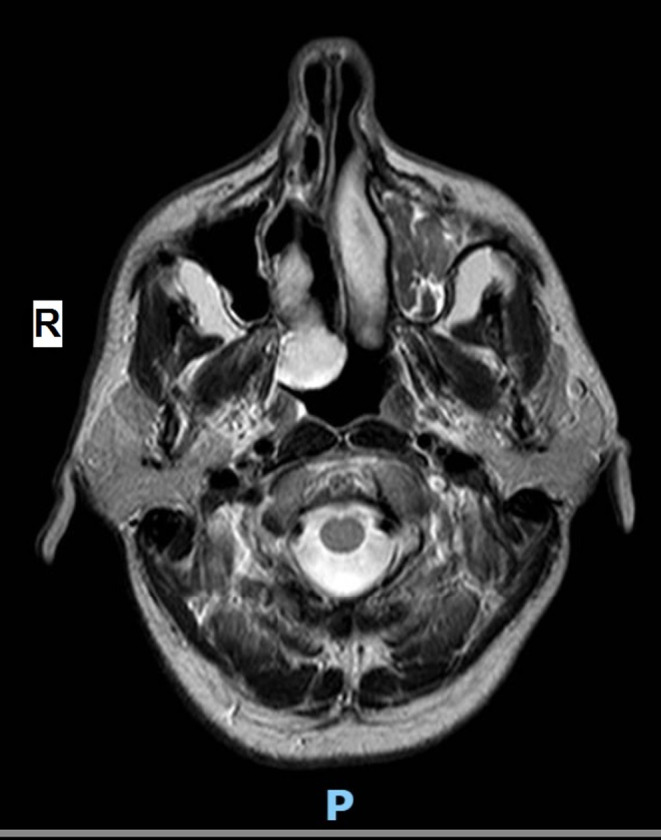
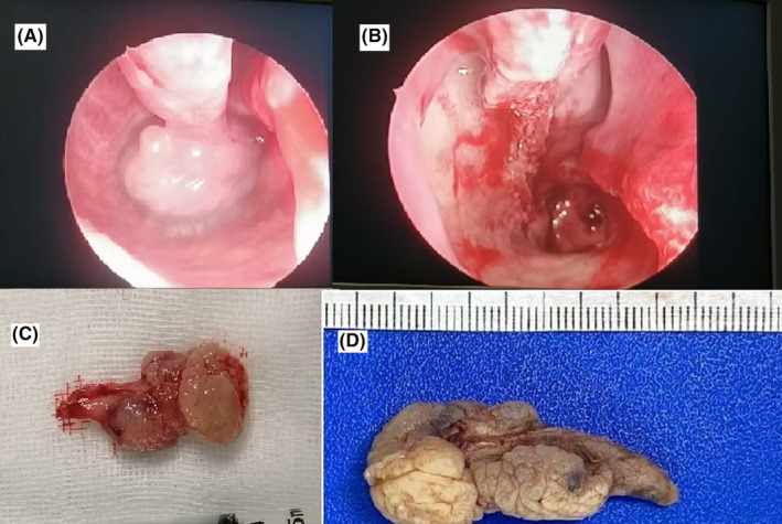
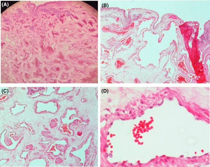
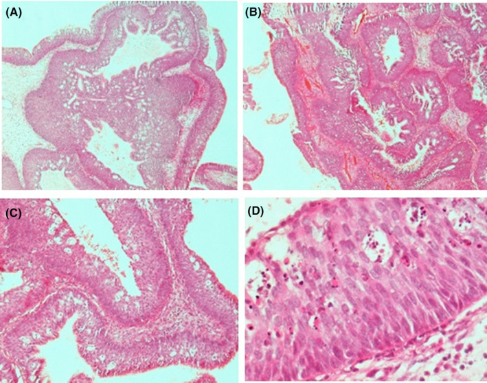
Hemangiomas are common lesions in medical practice, but those arising in the nasal cavity and/or paranasal sinuses are rare. Inverted papilloma is a rare benign tumor with a high incidence rate in both the nasal cavity and sinuses. The presence of both lesions in the same patient is even rarer. Here we present a case of a male patient with an unusual presentation and dual pathologies of cavernous hemangioma and inverted papilloma of the sinonasal tract that underwent endoscopic surgery and showed no recurrence of disease after a 2-year follow-up. The association of nasal cavernous hemangioma at one side and inverted papilloma at the other side is a very rare occasion that requires further studies and histopathology is the only diagnostic tool.
Keywords: hemangioma; histopathology; inverted papilloma; nasal polyp.
© 2023 The Authors. Clinical Case Reports published by John Wiley & Sons Ltd.
Conflict of interest statement
The authors declare no conflict of interest.
Figures
Similar articles
-
Nasal Malignant Melanoma With an Inverted Papilloma in the Contralateral Nasal Cavity: A Case Report.Ear Nose Throat J. 2023 Jun 8:1455613231179692. doi: 10.1177/01455613231179692. Online ahead of print. Ear Nose Throat J. 2023. PMID: 37291873
-
Challenging Nasal Pathologies: An Inverted Nasal Papilloma Case Series Illustrating Diagnostic Challenges and Management Strategies.Cureus. 2024 Aug 10;16(8):e66577. doi: 10.7759/cureus.66577. eCollection 2024 Aug. Cureus. 2024. PMID: 39252714 Free PMC article.
-
Role of radiological corroboration in a locally aggressive inverted papilloma: a case report.Ann Med Surg (Lond). 2023 Aug 14;85(10):5171-5175. doi: 10.1097/MS9.0000000000001193. eCollection 2023 Oct. Ann Med Surg (Lond). 2023. PMID: 37811095 Free PMC article.
-
Inverted papilloma with bilateral origin. A case report and review of the literature.Ann Ital Chir. 2022 Oct 5;11:S2239253X22038294. Ann Ital Chir. 2022. PMID: 36259423 Review.
-
Basaloid squamous cell carcinoma arising in an inverted papilloma in the nasal cavity: A case report and review.Auris Nasus Larynx. 2017 Oct;44(5):624-628. doi: 10.1016/j.anl.2016.09.005. Epub 2016 Oct 7. Auris Nasus Larynx. 2017. PMID: 27720480 Review.
Cited by
-
Primary Tuberculosis of Tonsils: Interesting Case Detected During the Histopathological Examination.Cureus. 2024 May 3;16(5):e59616. doi: 10.7759/cureus.59616. eCollection 2024 May. Cureus. 2024. PMID: 38832186 Free PMC article.
References
-
- Euteneuer S, Sudhoff H, Bernal‐Sprekelsen M, Theegarten D, Dazert S. Malignomas of the nasal cavity and the paranasal sinuses:clinical characteristics, therapy and prognosis of differenttumour types. Laryngorhinootologie. 2004;83(1):33‐39. - PubMed
-
- Hasan A, Nady M, Ibrahim A, et al. The utility of clinico‐pathological correlation of sinonasal masses in a tertiary hospital. J Evolution Med Dent Sci. 2021;10(10):679‐683.
-
- Sahin M. Chapter 589. Neurocutaneous syndromes. In: Behrman RE et al., eds. Nelson textbook of pediatrics. 19th ed. W.B. Saunders Company; 2011:2046‐2053 e1.
-
- Thapa N. Diagnosis and treatment of sinonasal inverted papilloma. Nepalese J ENT Head Neck Surg. 2010;1:30‐33.
Publication types
LinkOut - more resources
Full Text Sources
