

A Rare Case of Isolated Right Ventricular Loeffler's Endocarditis in Primary Hypereosinophilic Syndrome
- PMID: 38161771
- PMCID: PMC10756316
- DOI: 10.4103/jcecho.jcecho_22_23
A Rare Case of Isolated Right Ventricular Loeffler's Endocarditis in Primary Hypereosinophilic Syndrome
Abstract
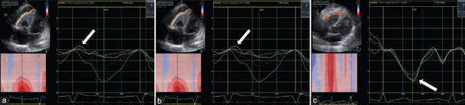
Hypereosinophilic syndrome (HES) is a systemic disorder with various manifestations, characterized by hypereosinophilia and caused by primary or secondary conditions. Loeffler's endocarditis (LE) represents a frequent cardiac manifestation of HES, caused by infiltration of the myocardium by eosinophilic cells, which determines endocardial damage, with subsequent inflammation, thrombosis, and fibrosis of either one or both ventricles. The diagnosis of cardiac involvement is based on a multimodality approach (i.e., two-dimensional transthoracic echocardiography [2D-TTE], speckle-tracking echocardiography [STE], and cardiac magnetic resonance [CMR]), with different findings depending on the stage of disease. STE may be useful in the initial phase when traditional imaging techniques may result negative, whereas CMR allows myocardial tissue characterization along with a better definition of the right ventricle. We present a rare case of LE with isolated right ventricular involvement in a patient with HES caused by chronic eosinophilic leukemia with constitutively activated fusion tyrosine kinase on chromosome 4q12, successfully treated with imatinib mesylate.
Keywords: Cardiac magnetic resonance; Loeffler’s endocarditis; multimodality imaging; speckle-tracking echocardiography.
Copyright: © 2023 Journal of Cardiovascular Echography.
Conflict of interest statement
There are no conflicts of interest.
Figures


References
-
- Salih M, Ibrahim R, Tirunagiri D, Al-Ani H, Ananthasubramaniam K. Loeffler's endocarditis and hypereosinophilic syndrome. Cardiol Rev. 2021;29:150–5. - PubMed
-
- Polito MV, Hagendorff A, Citro R, Prota C, Silverio A, De Angelis E, et al. Loeffler's endocarditis:An integrated multimodality approach. J Am Soc Echocardiogr. 2020;33:1427–41. - PubMed
-
- Carnero-Alcázar M, Reguillo-Lacruz F, O'Connor F, Rodríguez-Hernández E. Hypereosinophilic syndrome and myocardial fibrosis. Interact Cardiovasc Thorac Surg. 2008;7:928–30. - PubMed
-
- Beedupalli J, Modi K. Early-stage Loeffler's endocarditis with isolated right ventricular involvement:Management, long-term follow-up, and review of literature. Echocardiography. 2016;33:1422–7. - PubMed
Publication types
LinkOut - more resources
Full Text Sources
Research Materials