. 2023 Nov;18(6):100869.
doi: 10.1016/j.ajps.2023.100869. Epub 2023 Nov 28.

An infection-microenvironment-targeted and responsive peptide-drug nanosystem for sepsis emergency by suppressing infection and inflammation

Affiliations

An infection-microenvironment-targeted and responsive peptide-drug nanosystem for sepsis emergency by suppressing infection and inflammation

Wei He et al. Asian J Pharm Sci. 2023 Nov.

Abstract

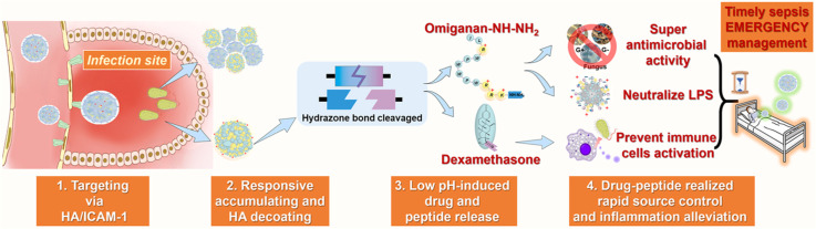
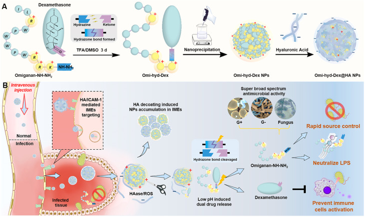
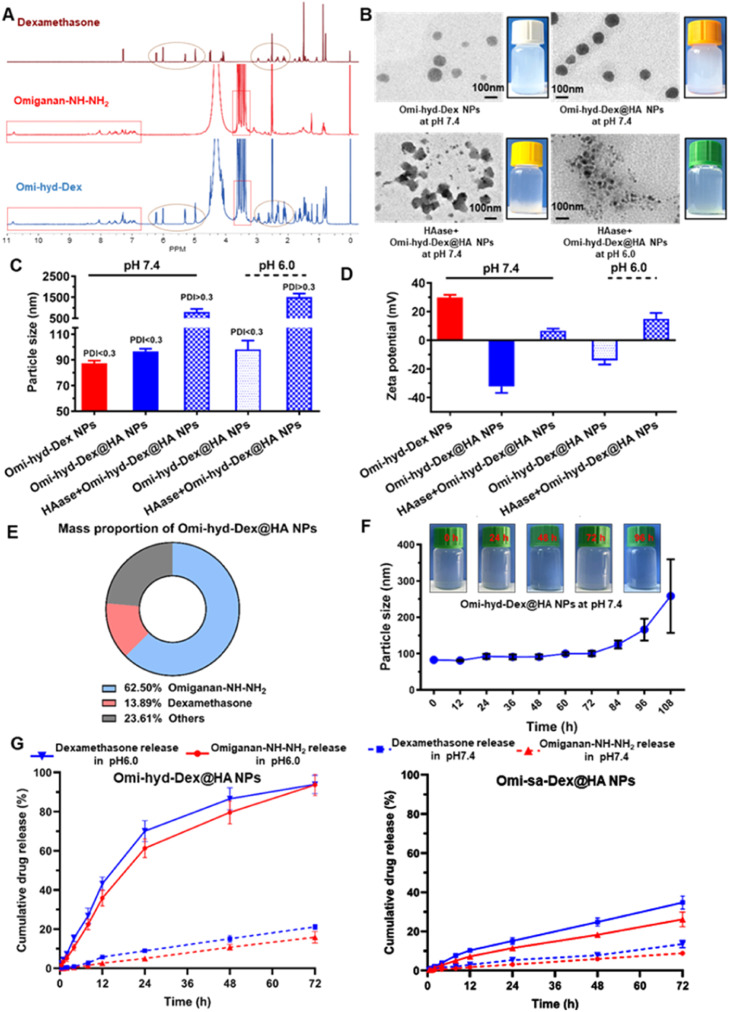
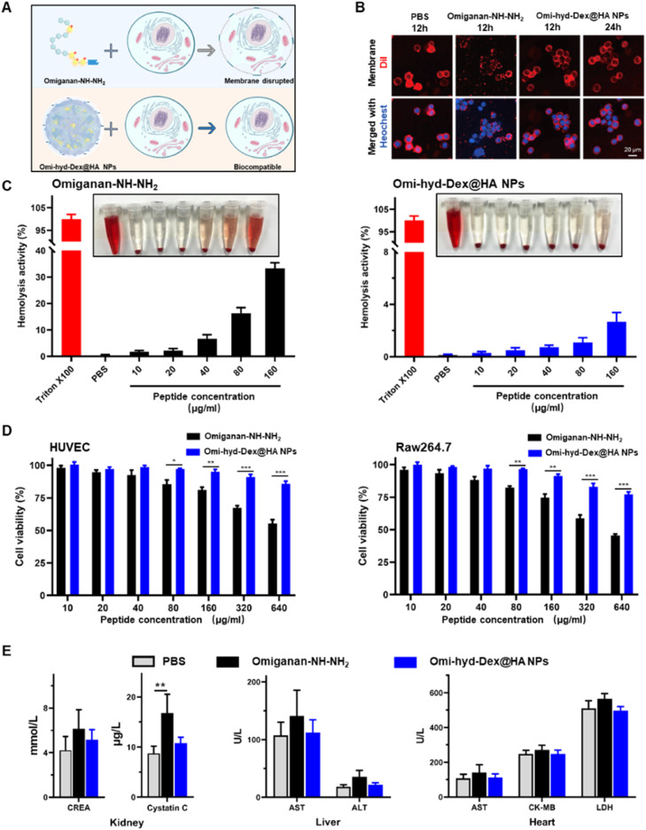
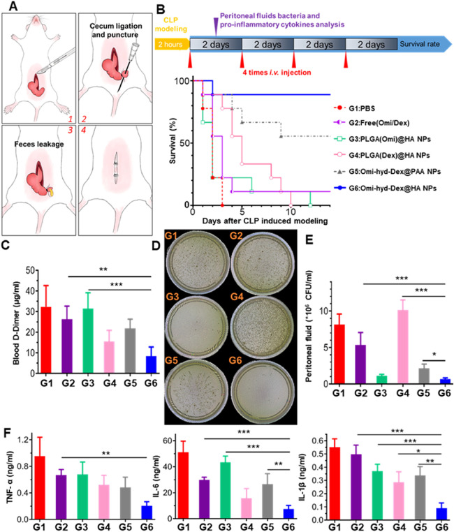
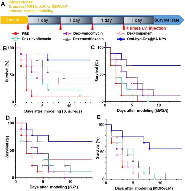
Sepsis is a life-threatening emergency that causes millions of deaths every year due to severe infection and inflammation. Nevertheless, current therapeutic regimens are inadequate to promptly address the vast diversity of potential pathogens. Omiganan, an antimicrobial peptide, has shown promise for neutralizing endotoxins and eliminating diverse pathogens. However, its clinical application is hindered by safety and stability concerns. Herein, we present a nanoscale drug delivery system (Omi-hyd-Dex@HA NPs) that selectively targets infectious microenvironments (IMEs) and responds to specific stimuli for efficient intervention in sepsis. The system consists of omiganan-dexamethasone conjugates linked by hydrazone bonds which self-assemble into nanoparticles coated with a hyaluronic acid (HA). The HA coating not only facilitates IMEs-targeting through interaction with intercellular-adhesion-molecule-1 on inflamed endotheliocytes, but also improves the biosafety of the nanosystem and enhances drug accumulation in primary infection sites triggered by hyaluronidase. The nanoparticles release dual drugs in IMEs through pH-sensitive cleavage of hydrazone bonds to eradicate pathogens and suppress inflammation. In multiple tissue infection and sepsis animal models, Omi-hyd-Dex@HA NPs exhibited rapid source control and comprehensive inflammation reduction, thereby preventing subsequent fatal complications and significantly improving survival outcomes. The bio-responsive and self-delivering nanosystem offers a promising strategy for systemic sepsis treatment in emergencies.

Keywords: Infectious microenvironments; Nanoscale drug delivery systems; Omiganan; Pathogens; Sepsis.


Conflict of interest statement

The authors report no conflicts of interest. The authors alone are responsible for the content and writing of this article.

Figures

Image, graphical abstract
Graphical abstract
Scheme 1
Scheme 1
Systemic treatment of sepsis by Omi-hyd-Dex@HA NPs. (A) The preparation process of Omi-hyd-Dex@HA NPs. Omi-hyd-Dex NPs were formed by PLGA-assisted self-assembly of hydrazone-linked peptide-drug conjugates (Omi-hyd-Dex). (B) Schematic illustration of the smart dual-drug delivery approach for systemic treatment of sepsis by Omi-hyd-Dex@HA NPs. The NPs targeted the IMEs and responsively released AMP (omiganan) and anti-inflammatory agent (Dex) for the emergent treatment of sepsis caused by multiple pathogens.
Fig 1
Fig. 1
Preparation and characterization of Omi-hyd-Dex@HA NPs. (A) 1H NMR spectra of Dex, Omiganan-NH—NH2 and Omi-hyd-Dex in DMSO‑d6. (B) TEM images (scale bar 100 nm) and photographs of Omi-hyd-Dex NPs and Omi-hyd-Dex@HA NPs in different conditions. NPs (0.3 mg/ml) were suspended in buffers (pH 7.4 and 6.0) with or without HAase (150 U/ml) for 2 h at 37 °C. (C) Particle size (hydrodynamic diameter) of NPs (0.05 mg/ml) in buffer (pH 7.4 and 6.0) measured by DLS and their (D) surface zeta potential. (E) Mass proportion of Omi-hyd-Dex@HA NPs. (F) Particle size changes of Omi-hyd-Dex@HA NPs (0.05 mg/ml) during 108-h storage and the representative images of Omi-hyd-Dex@HA NPs at certain time points. (G) In vitro release profiles of Dex and Omiganan-NH—NH2 from Omi-hyd-Dex@HA NPs or Omi-sa-Dex@HA NPs in PBS (pH 7.4 or 6.0).
Fig 2
Fig. 2
Biocompatibility of Omi-hyd-Dex@HA NPs in vitro and in vivo. (A) Schematic illustration of Omi-hyd-Dex@HA NPs avoiding the membrane disruption effect of Omiganan-NH—NH2. (B) Cell membrane staining with DiI (red) after Raw264.7 macrophage cells treated with PBS, Omiganan-NH—NH2, or Omi-hyd-Dex@HA NPs for 12–24 h. The nuclei were stained with Hoechst 33342 (blue). (C) Hemolysis assay of Omiganan-NH—NH2 and Omi-hyd-Dex@HA NPs. (D) The cytotoxicity of Omi-hyd-Dex@HA NPs in HUVEC and Raw264.7 cells. (E) The sera from the healthy mice that received treatment were biochemically analyzed. The mice (n = 3) were intravenously injected with PBS, Omiganan-NH—NH2, or Omi-hyd-Dex@HA NPs (10 mg/kg, peptide equivalent; four times; at 2-d intervals) and blood was drawn the day after the last injection. CREA, creatinine; AST, aspartate aminotransferase; ALT, alanine aminotransferase; CK-MB, creatine kinase-MB; LDH, lactic acid dehydrogenase. Data are shown as mean ± SD, *P < 0.05, **P < 0.01, ***P < 0.001, N.S., not significant.
Fig 3
Fig. 3
Omi-hyd-Dex@HA NPs targeting and penetrating inflamed endothelium cells by ICAM-1 recognition and accumulating in bacterial infectious tissues. (A) Confocal laser scanning microscope images showed the ICAM-1/HA-mediated interaction between NPs (FITC-labeled; green) and TNF-α-activated HUVECs. The nuclei were stained with Hoechst 33342 (blue). (1) Cells treated with Omi-hyd-Dex@HA NPs without TNF-α activation; (2) TNF-α-activated cells treated with Omi-hyd-Dex@PAA NPs; (3) TNF-α-activated cells treated with Omi-hyd-Dex@HA NPs; (4) TNF-α-activated cells treated with Omi-hyd-Dex@HA NPs after CD44 blocking-up; (5) TNF-α-activated cells treated with Omi-hyd-Dex@HA NPs after ICAM-1 blocking-up; (6) TNF-α-activated cells treated with Omi-hyd-Dex@HA NPs after free HA competitive inhibition. (B) Mean FITC fluorescence intensity in each group of HUVECs, which was calculated by adding the total FITC fluorescence intensity in each image together and dividing by the number of Hoechst 33342 labeled cells. (C) Omi-hyd-Dex@HA NPs penetrated each well of HUVECs after 24-h incubation. (D) Omi-hyd-Dex@HA NPs accumulated in K.P.-infected (tracheal injection) lung tissue. One dose of PBS or NPs (5 mg/kg; DiR-labeled) was administrated by i.v. injection to K.P.-induced pneumonia model mice (n = 3), and the major organs were taken out for IVIS imaging after 6 or 24 h. (E) Omi-hyd-Dex@HA NPs accumulated in S. aureus-infected footpad (left hind limb). One dose of PBS or NPs (5 mg/kg; DiR-labeled) was administrated by i.v. injection on S. aureus-induced footpad infection model mice (n = 3) and mice were imaged by IVIS after 6 or 24 h. (F) The mean fluorescence intensity per mm2 in major organs after i.v. injection for 6 or 24 h. K.P.-infected tissue: lung. (G) The mean fluorescence intensity per mm2 in hind limbs 6 or 24 h after i.v. injection. S. aureus-infected tissue: left hind limb. Data are shown as mean ± SD, *P < 0.05, ***P < 0.001, N.S., not significant.
Fig 4
Fig. 4
Omi-hyd-Dex@HA NPs retained the antibacterial activity of Omiganan-NH—NH2. (A) Representative SEM images of K.P. after treatment with PBS, Omiganan-NH—NH2 or Omi-hyd-Dex@HA NPs (2× MIC) for 4 h. (B-C) LIVE/DEAD stained K.P. under confocal laser scanning microscope. Red for PI-stained nucleoplasm, green for SYTO 9-stained bacterial membrane. Bacteria were treated with PBS, omiganan, or Omi-hyd-Dex@HA NPs (2× MIC) for 1 h, followed by staining with SYTO 9 (live and dead bacteria) and PI (dead bacteria). PI-positive bacteria were counted. N.S., not significant. (D-E) Time-kill kinetics assay of Omi-hyd-Dex@HA NPs, omiganan, or Gentamicin against K.P. and MDR-K.P. at different concentrations. Gentamicin was used as a positive control.
Fig 5
Fig. 5
The LPS neutralization and dual anti-inflammatory activity of Omi-hyd-Dex@HA NPs in vitro. (A) Schematic illustration of the dual anti-inflammatory function of Omi-hyd-Dex@HA NPs. (B-C) The released omiganan from Omi-hyd-Dex@HA NPs altered the particle state of LPS aggregation. The particle size and surface zeta potential were determined by the Malvern DLS device with an electrode probe. (D-E) E-selectin expression in mouse primary microvascular endothelial cells. LPS (100 ng/ml) with Omi-hyd-Dex@HA NPs or free drugs were mixed in DMSO solution and added into cell culture media for 24 h. (Omi-hyd-Dex@HA NPs 10 µg/ml, equivalent to Omiganan-NH—NH2 6.25 µg/ml or Dex 1.389 µg/ml). Vehicle (DMSO)-treated cells were employed as a negative control. (F) TNF-α and (G) NO production by LPS-challenged RAW264.7 macrophages after administration of Omi-hyd-Dex@HA NPs, Dex or Omiganan-NH—NH2. Data are shown as mean ± SD, *P < 0.05, **P < 0.01, ***P < 0.001, N.S., not significant. (1.44 mg of Omi-hyd-Dex@HA NPs contains 0.9 mg of Omiganan-NH—NH2 and 0.2 mg of Dex; LPS 100 ng/ml).
Fig 6
Fig. 6
The comprehensive anti-inflammation therapeutic efficacy of Omi-hyd-Dex@HA NPs in vivo. (A) Experiment timeline and survival percentages (n = 9) of lethal dose LPS-induced sepsis model mice receiving treatments. Sepsis mice treated with PBS were used as a negative control. (B) Neutrophils number, (C-E) pro-inflammatory cytokines concentrations, and (F) total protein mass quantification in peritoneal fluid collected from sepsis mice receiving various treatments (n = 3). (G-H) Blood white cells number and body temperature change 1 d after treatment. Compared with body temperature before modeling. Data are shown as mean ± SD, *P < 0.05, **P < 0.01, ***P < 0.001, N.S., not significant.
Fig 7
Fig. 7
Rounds of Omi-hyd-Dex@HA NPs treatment effectively controlled CLP-induced polymicrobial sepsis and reduced bacterial proliferation, d-Dimer level, and pro-inflammatory cytokines production. (A) Schematics of CLP modeling process. About 30% of the full length of the cecum was ligated and punctured with a 26 G needle for feces leakage. (B) Experiment timeline and survival percentages (n = 9) of CLP model mice receiving treatments. (C) Blood serum d-Dimer detection (n = 3) on Day 1 after treatment. (D) Representative photograph and (E) bacterial colonies number of peritoneal fluid inoculated on BHI agar plates in aerobic condition for 18 h. (F) Pro-inflammatory cytokines concentrations (n = 3) in peritoneal fluid of the mice receiving treatment. Data are shown as mean ± SD, *P < 0.05, **P < 0.01, ***P < 0.001, N.S., not significant.
Fig 8
Fig. 8
Simulation of Omi-hyd-Dex@HA NPs-based emergency therapeutic regimen against sepsis induced by four kinds of clinically isolated pathogenic bacteria. (A) Experiment timeline and legend name of each group. Mice were given intraperitoneal injections of four kinds of clinically isolated pathogenic bacteria for sepsis modeling. Mice with sepsis treated with PBS were used as a negative control. Drugs were administered once 1 d by i.v. injection for 4 times after modeling. (B-E) Survival percentages (n = 9 per group) of different bacteria-induced sepsis mice receiving treatments within 14 d

Similar articles

Cited by

References

    1. Cecconi M., Evans L., Levy M., Rhodes A. Sepsis and septic shock. Lancet. 2018;392(10141):75–87. - PubMed
    1. Hotchkiss R.S., Immunology Sherwood ER. Getting sepsis therapy right. Science. 2015;347(6227):1201–1202. - PMC - PubMed
    1. Rudd K.E., Johnson S.C., Agesa K.M., Shackelford K.A., Tsoi D., Kievlan D.R., et al. Global, regional, and national sepsis incidence and mortality, 1990-2017: analysis for the global burden of disease study. Lancet. 2020;395(10219):200–211. - PMC - PubMed
    1. Reitz K.M., Kennedy J., Li S.R., Handzel R., Tonetti D.A., Neal M.D., et al. Association between time to source control in sepsis and 90-day mortality. JAMA Surg. 2022;157(9):817–826. - PMC - PubMed
    1. Yuk S.A., Sanchez-Rodriguez D.A., Tsifansky M.D., Yeo Y. Recent advances in nanomedicine for sepsis treatment. Ther Deliv. 2018;9(6):435–450. - PMC - PubMed

LinkOut - more resources