

Causal link between prefrontal cortex and EEG microstates: evidence from patients with prefrontal lesion
- PMID: 38161794
- PMCID: PMC10757643
- DOI: 10.3389/fnins.2023.1306120
Causal link between prefrontal cortex and EEG microstates: evidence from patients with prefrontal lesion
Abstract
Introduction: At present, elucidating the cortical origin of EEG microstates is a research hotspot in the field of EEG. Previous studies have suggested that the prefrontal cortex is closely related to EEG microstate C and D, but whether there is a causal link between the prefrontal cortex and microstate C or D remains unclear.
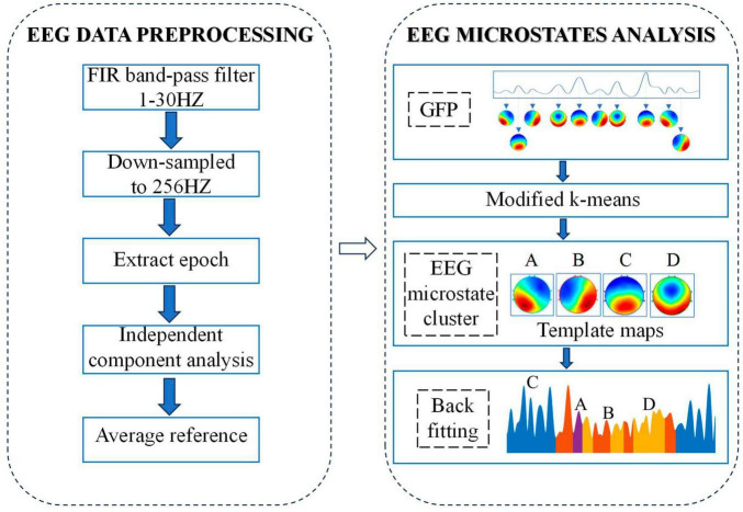
Methods: In this study, pretrial EEG data were collected from ten patients with prefrontal lesions (mainly located in inferior and middle frontal gyrus) and fourteen matched healthy controls, and EEG microstate analysis was applied.
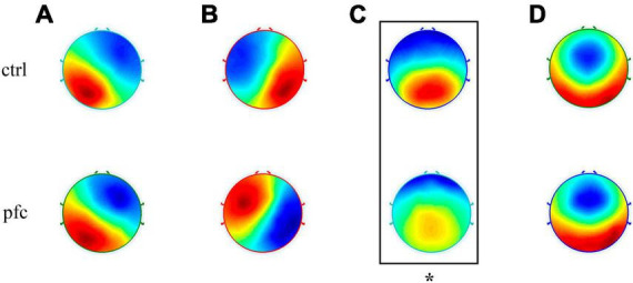
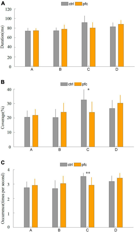
Results: Our results showed that four classical EEG microstate topographies were obtained in both groups, but microstate C topography in patient group was obviously abnormal. Compared to healthy controls, the average coverage and occurrence of microstate C significantly reduced. In addition, the transition probability from microstate A to C and from microstate B to C in patient group was significantly lower than those of healthy controls.
Discussion: The above results demonstrated that the damage of prefrontal cortex especially inferior and middle frontal gyrus could lead to abnormalities in the spatial distribution and temporal dynamics of microstate C not D, showing that there is a causal link between the inferior and middle frontal gyrus and the microstate C. The significance of our findings lies in providing new evidence for elucidating the cortical origin of microstate C.
Keywords: EEG; causal link; microstate; prefrontal cortex; prefrontal lesion.
Copyright © 2023 Zhao, Ran, Lv, Wang, Qiu, Wang, Xu, Guo, Gao, Mu and Yu.
Conflict of interest statement
The authors declare that the research was conducted in the absence of any commercial or financial relationships that could be construed as a potential conflict of interest.
Figures
Similar articles
-
The EEG microstate topography is predominantly determined by intracortical sources in the alpha band.Neuroimage. 2017 Nov 15;162:353-361. doi: 10.1016/j.neuroimage.2017.08.058. Epub 2017 Aug 25. Neuroimage. 2017. PMID: 28847493
-
EEG microstates as markers of major depressive disorder and predictors of response to SSRIs therapy.Prog Neuropsychopharmacol Biol Psychiatry. 2022 Jun 8;116:110514. doi: 10.1016/j.pnpbp.2022.110514. Epub 2022 Jan 24. Prog Neuropsychopharmacol Biol Psychiatry. 2022. PMID: 35085607
-
EEG microstates are correlated with brain functional networks during slow-wave sleep.Neuroimage. 2020 Jul 15;215:116786. doi: 10.1016/j.neuroimage.2020.116786. Epub 2020 Apr 7. Neuroimage. 2020. PMID: 32276057
-
Neural mechanisms of spatial navigation in ASD and TD children: insights from EEG microstate and functional connectivity analysis.Front Psychiatry. 2025 Apr 4;16:1552233. doi: 10.3389/fpsyt.2025.1552233. eCollection 2025. Front Psychiatry. 2025. PMID: 40256159 Free PMC article.
-
Distinct features of EEG microstates in autism spectrum disorder revealed by meta-analysis: the contribution of individual age to heterogeneity across studies.Front Psychiatry. 2025 Apr 22;16:1531694. doi: 10.3389/fpsyt.2025.1531694. eCollection 2025. Front Psychiatry. 2025. PMID: 40330653 Free PMC article.
Cited by
-
Abnormal nonlinear features of EEG microstate sequence in obsessive-compulsive disorder.BMC Psychiatry. 2024 Dec 4;24(1):881. doi: 10.1186/s12888-024-06334-6. BMC Psychiatry. 2024. PMID: 39627734 Free PMC article.
-
Classification of left and right-hand motor imagery in acute stroke patients using EEG microstate.J Neuroeng Rehabil. 2025 Jun 18;22(1):137. doi: 10.1186/s12984-025-01668-y. J Neuroeng Rehabil. 2025. PMID: 40533772 Free PMC article.
-
EEG-Based Analysis of Neural Responses to Sweeteners: Effects of Type and Concentration.Foods. 2025 Jul 14;14(14):2460. doi: 10.3390/foods14142460. Foods. 2025. PMID: 40724281 Free PMC article.
References
LinkOut - more resources
Full Text Sources