

Thalamic functional connectivity and sensorimotor processing in neurodevelopmental disorders
- PMID: 38161799
- PMCID: PMC10755010
- DOI: 10.3389/fnins.2023.1279909
Thalamic functional connectivity and sensorimotor processing in neurodevelopmental disorders
Abstract
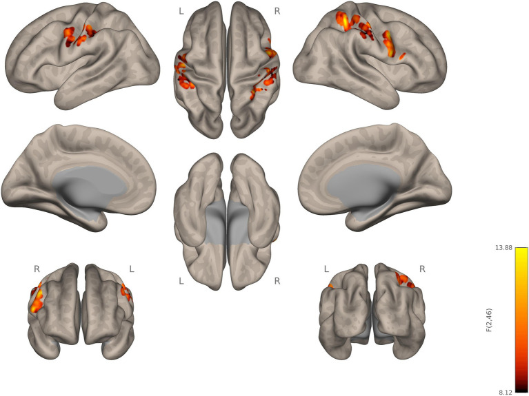
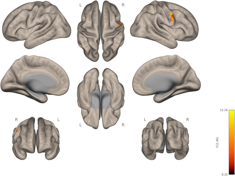
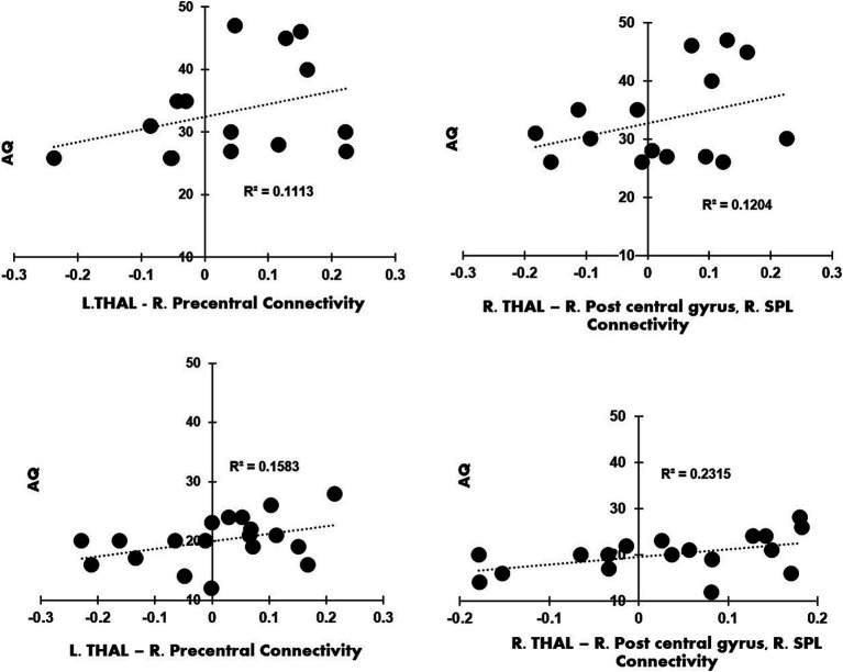
One of the earliest neurobiological findings in autism has been the differences in the thalamocortical pathway connectivity, suggesting the vital role thalamus plays in human experience. The present functional MRI study investigated resting-state functional connectivity of the thalamus in 49 (autistic, ADHD, and neurotypical) young adults. All participants underwent structural MRI and eyes-open resting state functional MRI scans. After preprocessing the imaging data using Conn's connectivity toolbox, a seed-based functional connectivity analysis was conducted using bilateral thalamus as primary seeds. Autistic participants showed stronger thalamic connectivity, relative to ADHD and neurotypical participants, between the right thalamus and right precentral gyrus, right pars opercularis-BA44, right postcentral gyrus, and the right superior parietal lobule (RSPL). Autistic participants also showed significantly increased connectivity between the left thalamus and the right precentral gyrus. Furthermore, regression analyses revealed a significant relationship between autistic traits and left thalamic-precentral connectivity (R2 = 0.1113), as well as between autistic traits and right postcentral gyrus and RSPL connectivity (R2 = 0.1204) in autistic participants compared to ADHD. These findings provide significant insights into the role of thalamus in coordinating neural information processing and its alterations in neurodevelopmental disorders.
Keywords: ADHD; autism; connectivity; fMRI; resting-state; thalamus.
Copyright © 2023 Karavallil Achuthan, Stavrinos, Argueta, Vanderburgh, Holm and Kana.
Conflict of interest statement
The authors declare that the research was conducted in the absence of any commercial or financial relationships that could be construed as a potential conflict of interest. The author(s) declared that they were an editorial board member of Frontiers, at the time of submission. This had no impact on the peer review process and the final decision.
Figures
Similar articles
-
Impairments of thalamic resting-state functional connectivity in patients with chronic tinnitus.Eur J Radiol. 2015 Jul;84(7):1277-84. doi: 10.1016/j.ejrad.2015.04.006. Epub 2015 Apr 20. Eur J Radiol. 2015. PMID: 25935516
-
Alterations of Functional Connectivity in Autism and Attention-Deficit/Hyperactivity Disorder Revealed by Multi-Voxel Pattern Analysis.Brain Connect. 2023 Nov;13(9):528-540. doi: 10.1089/brain.2023.0006. Epub 2023 Aug 22. Brain Connect. 2023. PMID: 37522594
-
Altered thalamic functional connectivity and cerebral blood flow in insomnia disorder: a resting-state functional magnetic resonance imaging study.Clin Imaging. 2022 Aug;88:17-23. doi: 10.1016/j.clinimag.2022.04.015. Epub 2022 May 4. Clin Imaging. 2022. PMID: 35561588
-
Resting-state functional connectivity networks associated with fatigue in multiple sclerosis with early age onset.Mult Scler Relat Disord. 2019 Jun;31:101-105. doi: 10.1016/j.msard.2019.03.020. Epub 2019 Mar 30. Mult Scler Relat Disord. 2019. PMID: 30954931
-
Functional connectivity alternation of the thalamus in restless legs syndrome patients during the asymptomatic period: a resting-state connectivity study using functional magnetic resonance imaging.Sleep Med. 2014 Mar;15(3):289-94. doi: 10.1016/j.sleep.2013.09.030. Epub 2014 Jan 30. Sleep Med. 2014. PMID: 24555993
Cited by
-
Twinned neuroimaging analysis contributes to improving the classification of young people with autism spectrum disorder.Sci Rep. 2024 Aug 29;14(1):20120. doi: 10.1038/s41598-024-71174-z. Sci Rep. 2024. PMID: 39209988 Free PMC article.
-
Combination of motor, sensory and affective tasks in an EEG paradigm for children with developmental disabilities.MethodsX. 2024 Oct 10;13:102997. doi: 10.1016/j.mex.2024.102997. eCollection 2024 Dec. MethodsX. 2024. PMID: 39498122 Free PMC article.
References
-
- Ayub R., Sun K. L., Flores R. E., Lam V. T., Jo B., Saggar M., et al. . (2021). Thalamocortical connectivity is associated with autism symptoms in high-functioning adults with autism and typically developing adults. Transl. Psychiatry 11, 1–9. doi: 10.1038/s41398-021-01221-0, PMID: - DOI - PMC - PubMed
LinkOut - more resources
Full Text Sources
Miscellaneous