

Uncommon Presentation of Craniopharyngioma: A Case Report of Post-traumatic Intratumoral Hemorrhage Leading to Cerebral Infarct
- PMID: 38161815
- PMCID: PMC10756656
- DOI: 10.7759/cureus.49669
Uncommon Presentation of Craniopharyngioma: A Case Report of Post-traumatic Intratumoral Hemorrhage Leading to Cerebral Infarct
Abstract
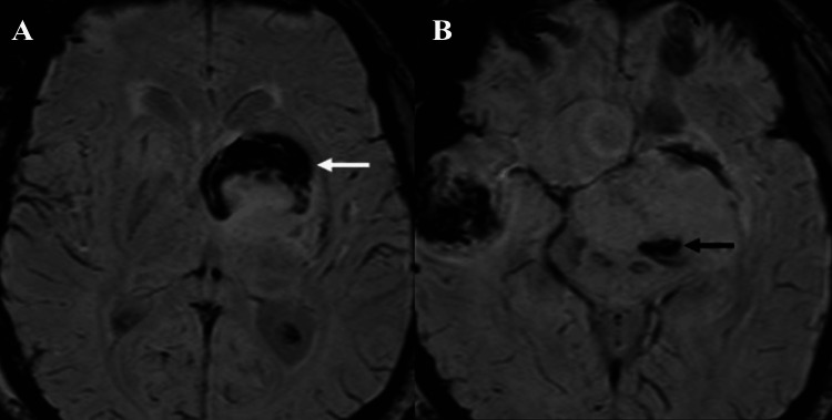
Craniopharyngiomas emanate from squamous cell remnants in the hypophyseal/pharyngeal duct region. This report details the unprecedented case of a 29-year-old male with adamantinomatous craniopharyngioma, who, following a motor vehicle collision (MVC), presented with post-traumatic intratumoral hemorrhage leading to acute basal ganglia infarct. The patient, previously subjected to subtotal resection, exhibited focal neurological deficits attributed to compression of lenticulostriate arteries due to the sudden increase in tumor volume. The patient, ineligible for thrombolysis or thrombectomy, was conservatively managed post-MVC. Subtotal resection occurred four months later. After one year, persistent right-sided weakness (2/5 motor power) remained, and the recommended stereotactic radiotherapy was declined by the patient. Notably, this instance represents the first documented case of post-traumatic intratumoral hemorrhage in adamantinomatous craniopharyngioma. This report distinguishes between adamantinomatous and papillary subtypes, noting their prevalence in different age groups. While these tumors commonly present with gradual vision changes, fatigue, and endocrine dysfunction, complications such as intra-tumoral hemorrhage remain rare. This report serves as an educational tool, shedding light on potential complications and urging increased vigilance in managing craniopharyngiomas.
Keywords: adamantinomatous craniopharyngioma; intratumoral hemorrhage; lenticulostriate artery; neuro radiology; supra sellar mass.
Copyright © 2023, Hegde et al.
Conflict of interest statement
The authors have declared that no competing interests exist.
Figures


Similar articles
-
A rare case of intratumoral hemorrhage in a young adult with adamantinomatous craniopharyngioma.Radiol Case Rep. 2024 Nov 14;20(1):761-766. doi: 10.1016/j.radcr.2024.10.044. eCollection 2025 Jan. Radiol Case Rep. 2024. PMID: 39624710 Free PMC article.
-
A Rare Case of Adamantinomatous Craniopharyngioma in an Adult.Cureus. 2022 Oct 6;14(10):e30000. doi: 10.7759/cureus.30000. eCollection 2022 Oct. Cureus. 2022. PMID: 36381754 Free PMC article.
-
Craniopharyngioma in Pediatrics and Adults.Adv Exp Med Biol. 2023;1405:299-329. doi: 10.1007/978-3-031-23705-8_11. Adv Exp Med Biol. 2023. PMID: 37452943
-
Suprasellar squamous papillary craniopharyngioma: a case report.Optometry. 2001 May;72(5):299-308. Optometry. 2001. PMID: 11394840 Review.
-
First evidence of anti-VEGF efficacy in an adult case of adamantinomatous craniopharyngioma: Case report and illustrative review.Ann Endocrinol (Paris). 2023 Dec;84(6):727-733. doi: 10.1016/j.ando.2023.10.003. Epub 2023 Oct 19. Ann Endocrinol (Paris). 2023. PMID: 37865272 Review.
References
-
- Craniopharyngioma and other cystic epithelial lesions of the sellar region: a review of clinical, imaging, and histopathological relationships. Zada G, Lin N, Ojerholm E, Ramkissoon S, Laws ER. Neurosurg Focus. 2010;28:0. - PubMed
-
- Imaging of craniopharyngioma. Curran JG, O'Connor E. Childs Nerv Syst. 2005;21:635–639. - PubMed
-
- Craniopharyngioma: a comparison of tumor control with various treatment strategies. Yang I, Sughrue ME, Rutkowski MJ, et al. Neurosurg Focus. 2010;28:0. - PubMed
-
- Significance of hemorrhage into brain tumors: clinicopathological study. Kondziolka D, Bernstein M, Resch L, Tator CH, Fleming JF, Vanderlinden RG, Schutz H. J Neurosurg. 1987;67:852–857. - PubMed
Publication types
LinkOut - more resources
Full Text Sources