Multiple Myeloma Treatment Challenges: A Case Report of Vertebral Artery Pseudoaneurysm Complicating Occipitocervical Arthrodesis and a Review of the Literature
- PMID: 38161862
- PMCID: PMC10757504
- DOI: 10.7759/cureus.49716
Multiple Myeloma Treatment Challenges: A Case Report of Vertebral Artery Pseudoaneurysm Complicating Occipitocervical Arthrodesis and a Review of the Literature
Abstract
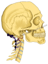
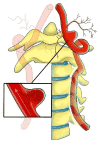
Multiple myeloma is a hematological neoplasm that frequently affects the spinal column. Less than a fifth of this vertebral involvement corresponds to the cervical spine and cranio-cervical junction. When there is instability or neurological involvement due to compression or deformity, approaches for anterior decompression and occipitocervical stabilization are required. The correct managment of vertebral artery aneurysm associated with occipitocervical arthrodesis requires extensive knowledge of anatomy and pathology. We present a case of a vertebral pseudoaneurysm that occurred late after the resection of a C1-C2 vertebral body multiple myeloma lesion managed with endonasal endoscopic approach and posterior occipitocervical arthrodesis as well as a systematic review of the related literature. The patient recovered well, without major neurological deficits.
Keywords: arthrodesis; cervical spine; endovascular treatment; multiple myeloma; spine oncology; surgical complication.
Copyright © 2023, Reyes Soto et al.
Conflict of interest statement
The authors have declared that no competing interests exist.
Figures


Similar articles
-
Pediatric cervical kyphosis in the MRI era (1984-2008) with long-term follow up: literature review.Childs Nerv Syst. 2022 Feb;38(2):361-377. doi: 10.1007/s00381-021-05409-z. Epub 2021 Nov 22. Childs Nerv Syst. 2022. PMID: 34806157 Review.
-
Pediatric occipitocervical fusion: long-term radiographic changes in curvature, growth, and alignment.J Neurosurg Pediatr. 2016 Nov;18(5):644-652. doi: 10.3171/2016.4.PEDS15567. Epub 2016 Jul 29. J Neurosurg Pediatr. 2016. PMID: 27472669
-
C2-C3 Anterior Cervical Arthrodesis in the Treatment of Bow Hunter's Syndrome: Case Report and Review of the Literature.World Neurosurg. 2018 Oct;118:284-289. doi: 10.1016/j.wneu.2018.07.129. Epub 2018 Jul 24. World Neurosurg. 2018. PMID: 30053560 Review.
-
Assessment of the results of occipito-cervical stabilization in cranio-vertebral damage.Pol Merkur Lekarski. 2020 Aug 22;49(286):228-231. Pol Merkur Lekarski. 2020. PMID: 32827415
-
Balloon kyphoplasty and additional anterior odontoid screw fixation for treatment of unstable osteolytic lesions of the vertebral body C2: a case series.BMC Musculoskelet Disord. 2018 Jul 27;19(1):259. doi: 10.1186/s12891-018-2180-x. BMC Musculoskelet Disord. 2018. PMID: 30049274 Free PMC article.
Cited by
-
Pioneering Augmented and Mixed Reality in Cranial Surgery: The First Latin American Experience.Brain Sci. 2024 Oct 16;14(10):1025. doi: 10.3390/brainsci14101025. Brain Sci. 2024. PMID: 39452038 Free PMC article.
-
Latex vascular injection as method for enhanced neurosurgical training and skills.Front Surg. 2024 Feb 23;11:1366190. doi: 10.3389/fsurg.2024.1366190. eCollection 2024. Front Surg. 2024. PMID: 38464665 Free PMC article.
-
Correlation of Edema/Tumor Index With Histopathological Outcomes According to the WHO Classification of Cranial Tumors.Cureus. 2024 Nov 3;16(11):e72942. doi: 10.7759/cureus.72942. eCollection 2024 Nov. Cureus. 2024. PMID: 39634980 Free PMC article.
-
Intraoperative Ultrasound: An Old but Ever New Technology for a More Personalized Approach to Brain Tumor Surgery.Cureus. 2024 Jun 12;16(6):e62278. doi: 10.7759/cureus.62278. eCollection 2024 Jun. Cureus. 2024. PMID: 39006708 Free PMC article.
-
The Vertebrobasilar Trunk and Its Anatomical Variants: A Microsurgical Anatomical Study.Diagnostics (Basel). 2024 Mar 2;14(5):534. doi: 10.3390/diagnostics14050534. Diagnostics (Basel). 2024. PMID: 38473006 Free PMC article.
References
-
- Multiple myeloma of the cervical spine: treatment strategies for pain and spinal instability. Rao G, Ha CS, Chakrabarti I, Feiz-Erfan I, Mendel E, Rhines LD. J Neurosurg Spine. 2006;5:140–145. - PubMed
-
- Evolution of treatment for metastatic spine disease. Wu AS, Fourney DR. Neurosurg Clin N Am. 2004;15:401–411. - PubMed
-
- Managing the cervical spine in multiple myeloma patients. Cawley DT, Butler JS, Benton A, et al. Hematol Oncol. 2019;37:129–135. - PubMed
-
- Advances in the treatment of metastatic spine tumors: the future is not what it used to be. Laufer I, Bilsky MH. J Neurosurg Spine. 2019;30:299–307. - PubMed
Publication types
LinkOut - more resources
Full Text Sources
Miscellaneous
