

Expression of Gata Binding Protein 3 as a Prognostic Factor in Urogenital Lesions and Its Association With Morphology
- PMID: 38161907
- PMCID: PMC10755802
- DOI: 10.7759/cureus.49635
Expression of Gata Binding Protein 3 as a Prognostic Factor in Urogenital Lesions and Its Association With Morphology
Abstract
Background: Urogenital malignancies, encompassing urinary bladder cancer, prostate cancer, and renal cell carcinoma, pose significant diagnostic challenges due to overlapping histopathological features. GATA binding protein 3 (GATA3), a transcription factor associated with urothelial tissue, has shown promise as a potential diagnostic marker. This study aimed to investigate the incidence of these malignancies, explore GATA3's involvement in urothelial cancer (UC), and determine its role in distinguishing urogenital malignancies.
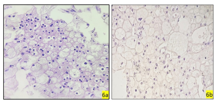
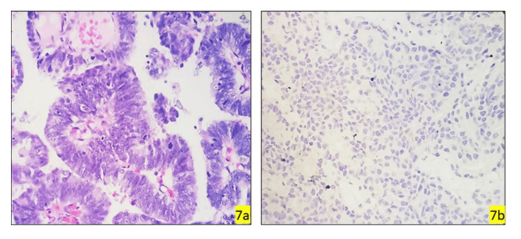
Materials and methods: A cross-sectional, retro-prospective, hospital-based study was conducted from May 2019 to April 2021. The surgical samples of patients who underwent transurethral resection of bladder tumour (TURBT), transurethral resection of the prostate (TURP), radical cystoprostatectomy, total and partial radical nephrectomy specimens during the study period were reviewed. Patients diagnosed with urinary bladder neoplasm and high-grade prostate neoplasm along with chromophobe, oncocytic, sarcomatoid variant and clear cell carcinoma, renal cell carcinoma were included. Immunohistochemical analysis of GATA3 expression was performed, with scoring based on nuclear staining intensity and percentage of tumor cells labeled.
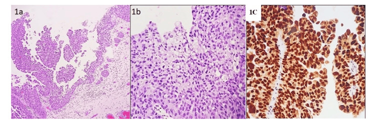
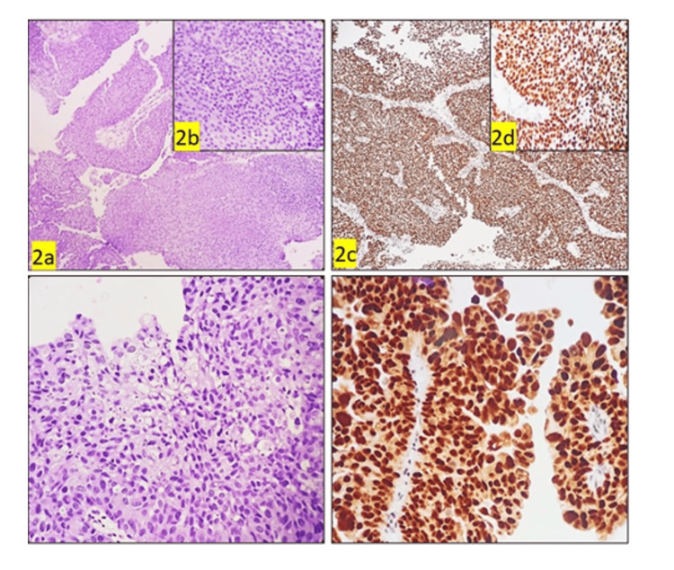
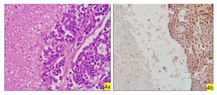
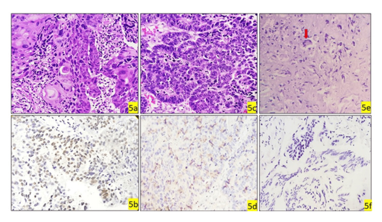
Results: The study included 64 patients, predominantly males over 60 years. Personal habits revealed a high prevalence of smoking (85.9%). The most prevalent symptom was hematuria (75.0%), followed by hematuria with urgency (20.3%). The most common site of lesion was posterolateral (31.3%). Urothelial cancer was the most common malignancy, primarily high-grade. Strong positive GATA3 expression was significantly associated with high-grade UC (p=0.01) and invasion (p=0.01). However, low-grade UC and papillary urothelial neoplasm of low malignant potential exhibited moderate GATA3 expression. GATA3 demonstrated potential for distinguishing UC from other histological types.
Conclusion: GATA3 expression correlates with high-grade urothelial cancer and invasive behavior, suggesting its utility as a diagnostic marker in challenging cases.
Keywords: gata3; histopathology; immunohistochemistry; prostate cancer; renal cell carcinoma; urothelial carcinoma.
Copyright © 2023, Govardhan et al.
Conflict of interest statement
The authors have declared that no competing interests exist.
Figures

Similar articles
-
Diagnostic utility of GATA3 immunohistochemical expression in urothelial carcinoma.Indian J Pathol Microbiol. 2019 Apr-Jun;62(2):244-250. doi: 10.4103/IJPM.IJPM_228_18. Indian J Pathol Microbiol. 2019. PMID: 30971548
-
Utility of GATA3 immunohistochemistry in differentiating urothelial carcinoma from prostate adenocarcinoma and squamous cell carcinomas of the uterine cervix, anus, and lung.Am J Surg Pathol. 2012 Oct;36(10):1472-6. doi: 10.1097/PAS.0b013e318260cde7. Am J Surg Pathol. 2012. PMID: 22982890 Free PMC article.
-
Utility of uroplakin II expression as a marker of urothelial carcinoma.Hum Pathol. 2015 Jan;46(1):58-64. doi: 10.1016/j.humpath.2014.09.007. Epub 2014 Oct 2. Hum Pathol. 2015. PMID: 25449628
-
Best practices recommendations in the application of immunohistochemistry in the prostate: report from the International Society of Urologic Pathology consensus conference.Am J Surg Pathol. 2014 Aug;38(8):e6-e19. doi: 10.1097/PAS.0000000000000238. Am J Surg Pathol. 2014. PMID: 25029122
-
Cytological and histological findings of upper tract mucinous urothelial carcinoma with clear cell component: A case report and review of literature.Diagn Cytopathol. 2022 May;50(5):E129-E135. doi: 10.1002/dc.24921. Epub 2021 Dec 27. Diagn Cytopathol. 2022. PMID: 34957705 Review.
References
-
- Urinary bladder cancer and its associated factors - an epidemiological overview. Mishra V, Balasubramaniam G. Indian J Med Sci. 2021;73:239–248.
LinkOut - more resources
Full Text Sources
Miscellaneous