

A Rare Case of Papillary Thyroid Carcinoma in the Thyroglossal Duct Cyst of a 14-Year-Old Female Patient With Left Thyroid Hemiagenesis
- PMID: 38161947
- PMCID: PMC10757459
- DOI: 10.7759/cureus.49712
A Rare Case of Papillary Thyroid Carcinoma in the Thyroglossal Duct Cyst of a 14-Year-Old Female Patient With Left Thyroid Hemiagenesis
Abstract
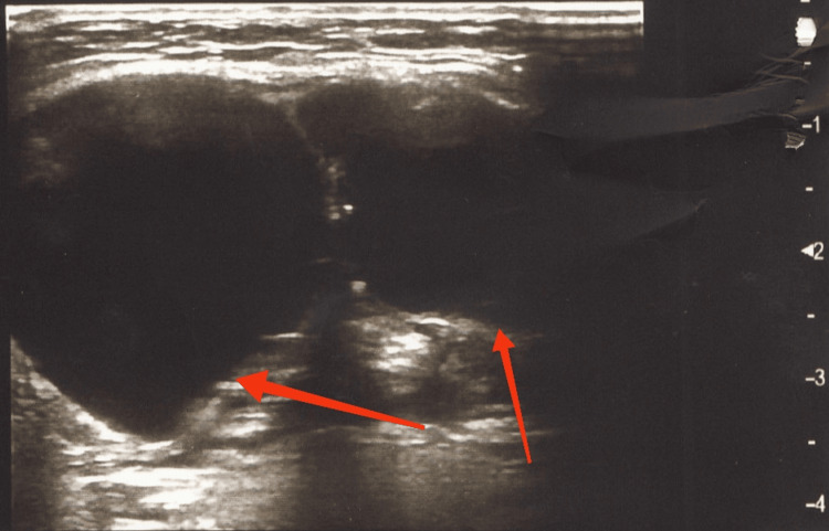
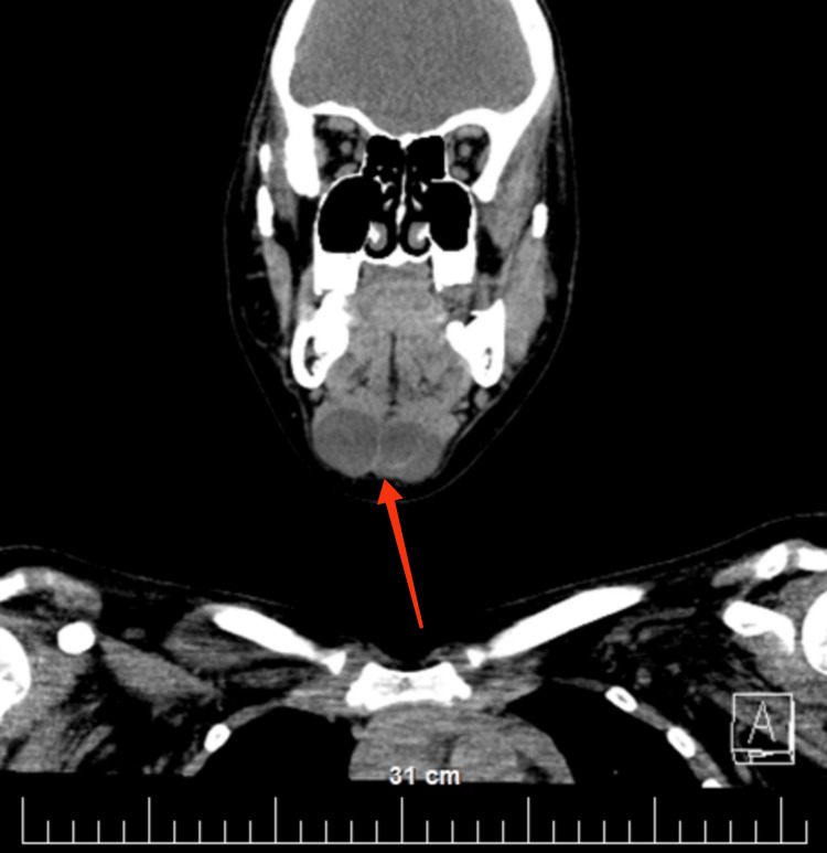
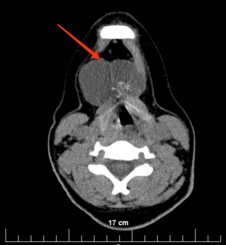
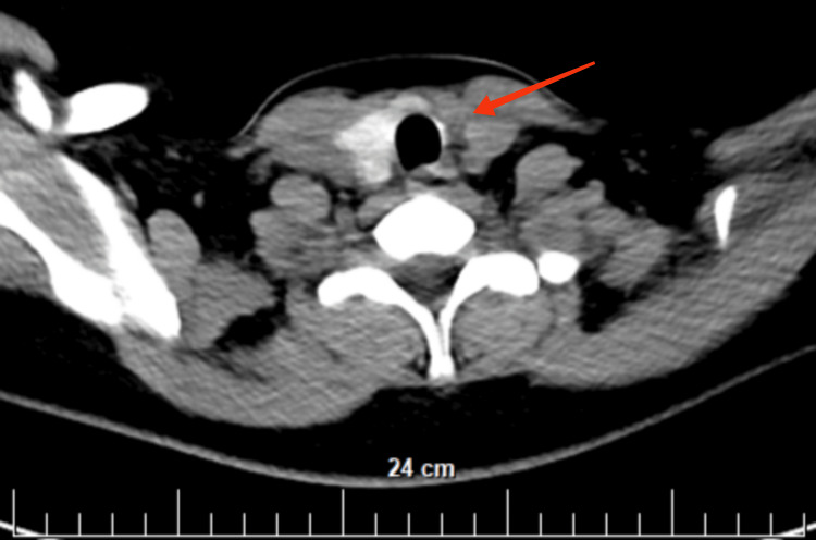
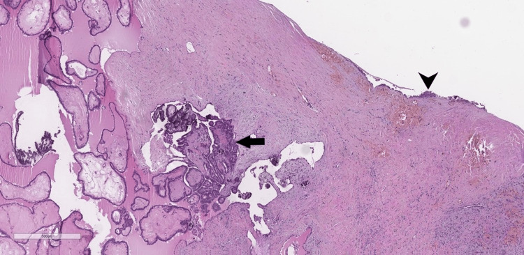
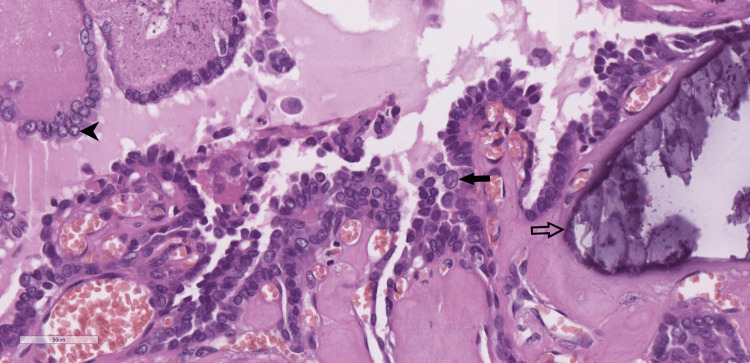
Congenital abnormalities in the development of the thyroglossal duct are a common pathology in the pediatric population. The exact frequency of hemiagenesis of the thyroid gland is not known because the condition is rarely manifested clinically and is almost always discovered incidentally. Papillary carcinoma of thyroglossal cysts is relatively uncommon, has a good prognosis if promptly detected and treated and occurs mainly in adults. The case we present here is an extremely rare occurrence: a patient with papillary thyroid carcinoma of the thyroglossal duct cyst and thyroglossal duct cyst carcinoma (TDCa). So far, only two such adult patients (women aged 24 and 35) have been described in the world medical literature. The patient we present is a 14-year-old female and is the first described adolescent with papillary carcinoma of the thyroglossal duct cyst and thyroid hemiagenesis (THA). The disease didn't have any clinical manifestations, and the patient was brought in by her parents to improve her aesthetic appearance. Neither the physical examination nor the radiological evaluation showed any signs of malignancy. The diagnosis was reached by our team only after the patoanatomical examination. In this patient's case, due to its early diagnosis, the spread of the disease was limited only to the borders of the thyroglossal duct cyst and the absence of regional and distant metastasis. Surgical removal led to a complete cure, without any postoperative data suggestive of residual disease. The functions of the thyroid gland in her case were not affected, despite her left-lobe agenesis, to which there are multiple proofs, namely the normal blood concentration of the examined thyroid markers: free triiodothyronine (FT3), free thyroxine (FT4), thyroglobulin (TG), thyroid stimulating hormone (TSH), anti-TG (thyroid antibody test (TAT)), anti-thyroid peroxidase (TPO) (microsomal antibody test (MAT)), and normal physical and psychological development.
Keywords: cancer in children; congenital anomalies; head and neck surgery; malignant cyst; maxillofacial surgery; median neck cyst; neck cancer; neck pathology; pediatric surgery; thyroid gland.
Copyright © 2023, Yankov et al.
Conflict of interest statement
The authors have declared that no competing interests exist.
Figures
Similar articles
-
Robot-assisted Sistrunk's operation, total thyroidectomy, and neck dissection via a transaxillary and retroauricular (TARA) approach in papillary carcinoma arising in thyroglossal duct cyst and thyroid gland.Ann Surg Oncol. 2012 Dec;19(13):4259-61. doi: 10.1245/s10434-012-2674-y. Epub 2012 Oct 16. Ann Surg Oncol. 2012. PMID: 23070784
-
Papillary carcinoma of a thyroglossal duct cyst in a patient with thyroid hemiagenesis: effectiveness of conservative surgical treatment.Endocr Pract. 2008 May-Jun;14(4):465-9. doi: 10.4158/EP.14.4.465. Endocr Pract. 2008. PMID: 18558601
-
Papillary Carcinoma of Thyroglossal Cyst with Thyroid Hemiagenesis: Is Conservative Surgical Management Enough?Indian J Otolaryngol Head Neck Surg. 2022 Oct;74(Suppl 2):1992-1995. doi: 10.1007/s12070-020-01951-5. Epub 2020 Jul 11. Indian J Otolaryngol Head Neck Surg. 2022. PMID: 36452739 Free PMC article.
-
Invasive Thyroglossal Duct Cyst Papillary Carcinoma: A Case Report and Review of the Literature.Am J Case Rep. 2018 Jun 28;19:757-762. doi: 10.12659/AJCR.907313. Am J Case Rep. 2018. PMID: 29950556 Free PMC article. Review.
-
Thyroglossal Duct Cyst Carcinomas in Pediatric Patients: Report of Two Cases with a Comprehensive Literature Review.Head Neck Pathol. 2017 Dec;11(4):442-449. doi: 10.1007/s12105-017-0807-0. Epub 2017 Mar 14. Head Neck Pathol. 2017. PMID: 28293858 Free PMC article. Review.
Cited by
-
The Origins and Development of Pre-emptive Dermatologic Anesthesia: A Systematic Review.Cureus. 2024 Mar 9;16(3):e55851. doi: 10.7759/cureus.55851. eCollection 2024 Mar. Cureus. 2024. PMID: 38590497 Free PMC article. Review.
-
An Unusual Mixed Thyroid Carcinoma: A Surgical and Pathological Rarity.Cureus. 2024 Apr 15;16(4):e58291. doi: 10.7759/cureus.58291. eCollection 2024 Apr. Cureus. 2024. PMID: 38752032 Free PMC article.
References
-
- Thyroid hemiagenesis from childhood to adulthood: review of literature and personal experience. De Sanctis V, Soliman AT, Di Maio S, Elsedfy H, Soliman NA, Elalaily R. https://pubmed.ncbi.nlm.nih.gov/27116848/ Pediatr Endocrinol Rev. 2016;13:612–619. - PubMed
-
- Prevalence of thyroid hemiagenesis: ultrasound screening in normal children. Shabana W, Delange F, Freson M, Osteaux M, De Schepper J. Eur J Pediatr. 2000;159:456–458. - PubMed
Publication types
LinkOut - more resources
Full Text Sources
Miscellaneous