. 2024 Jan-Feb;14(1):8-16.
doi: 10.1016/j.jobcr.2023.11.004. Epub 2023 Dec 8.

Putative effect of chronic renal failure on granular convoluted tubule of submandibular gland in male rats: Immunohistochemical and ultrastructural study

Affiliations

Putative effect of chronic renal failure on granular convoluted tubule of submandibular gland in male rats: Immunohistochemical and ultrastructural study

Wafaa Yahia Alghonemy et al. J Oral Biol Craniofac Res. 2024 Jan-Feb.

Abstract

Objective: This study aimed to investigate the ultrastructural and immunohistochemical changes in the granular convoluted tubule (GCT) of rodents' submandibular gland (SMG) upon theinduction of chronic renal failure.

Material and methods: Thirty young adult Sprague-Dawley rats were randomized into three groups: the Control group, rats received no intervention; the Sham group, rats underwent surgical incision without nephrectomy; the Experimental group, rats underwent surgical procedures to induce chronic renal failure. Afterward, SMG was examined for histological and ultrastructural changes and immunohistochemical staining for Renin.

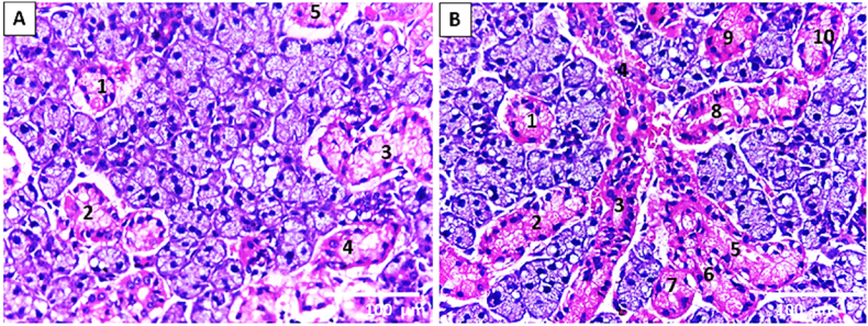
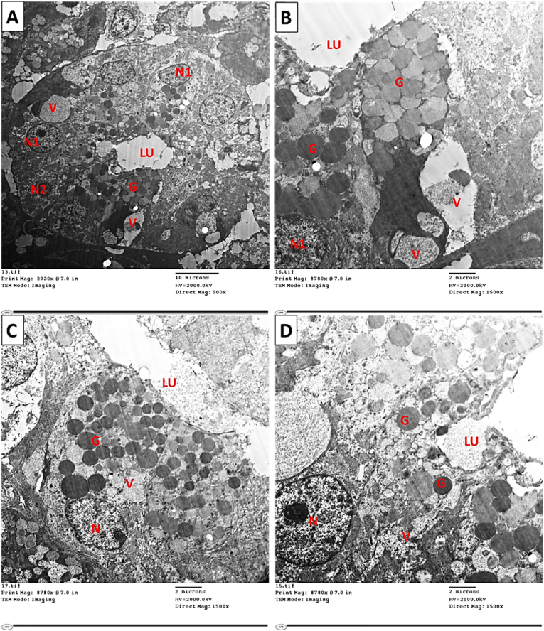
Results: Histologically, the experimental group demonstrated cytoplasmic vacuolization within the seromucous acini and ducts. Several GCTs were proliferating, whereas others exhibited degenerative changes in the form of disturbed cytoplasmic architecture. On the ultrastructural level, both acini and ductal segments showed degenerative changes Interestingly, immunohistochemical examination of the lining cells of GCT and intralobular ducts of the experimental group revealed the presence of Renin.

Conclusion: Renal failure induced histological, immunohistochemical, and ultrastructural variations within GCTs of SMG.

Keywords: Granular convoluted tubules; Renal failure; Renin; Submandibular salivary gland.


Conflict of interest statement

We wish to confirm that there are no known conflicts of interest associated with this publication and there has been no significant financial support for this work that could have influenced its outcome.

Figures

Image 1
Graphical abstract
Fig. 1
Fig. 1
Flowchart explaining the rat's randomization and the variables assessed throughout the study.
Fig. 2
Fig. 2
light microscopic image of rat SMG, as displayed on the Image Monitor. The GCT areas are outlined, and their number is recorded. (A) Control group as displayed on the image monitor. (B) Experimental group that reveals an obvious increase in GCTs (H&E stain. Orig. mag. X 400).
Fig. 3
Fig. 3
Light microscopic image of rat SMG illustrates (A) the control group's seromucous acini A and GCT. (BD) the seromucous acini and GCT of the experimental group. (B) It reveals the proliferation of GCT (black arrows) (embryonic-like branched structure). (C,D) It shows seromucous acini with cytoplasmic vacuolization (white arrows) together with the disturbing architecture of GCT (arrowhead). (H&E stain. Orig. mag. a, b x400; c x1000).
Fig. 4
Fig. 4
Light microscopic image of rat SMG illustrates (A) the almost normal seromucous acini and GCT of the sham group. (B,C) the seromucous acini and GCT of the experimental group reveal a marked proliferation of GCT. (D) It shows widely branched proliferated GCTs in between the seromucous acini (H&E stain. Orig. mag. A X1000; B, D X100; C X400).
Fig. 5
Fig. 5
Photomicrograph of immunolocalization of anti-renin antibodies in right SMG: (A , B) showing negative cellular and nuclear expression of anti-renin antibodies in both acini (yellow arrow) and ducts (red arrow) of control as well as sham groups, respectively. (C) showing lobes of SMG of experimental group after eight weeks of chronic Induction of renal failure (D) Higher magnification of black boxed area at (C) showing positive localization of cellular expression of anti-renin at the GCT and intralobular ducts lining cells (red arrows) with some nuclear immunohistochemical reaction. In addition, there is a negative expression of anti-renin in the gland acini (yellow arrow). (Anti-renin antibodies, A, B & D x 400 and C x 100).
Fig. 6
Fig. 6
Electron micrograph of SMG of control and sham groups showing (A,B) Normal pyramidal seromucous acinar cell with basal active nucleus (N) and normal appearance of RER occupying mainly the cell's basolateral part. The acinar cell appears loaded with electron-lucent secretory granules (G), which appear to merge. (C) SMG of the experimental group showing: Seromucous acinar cell with basal nucleus (N) and small cisterna of RER (arrow). The acinar cell appears loaded with both electron lucent and electron-dense secretory granules. Notice the presence of cytoplasmic vacuoles (V) of different sizes. Similarly, (D) Seromucous acinar cell with basal heterochromatic nucleus (N) and small cisterna of RER (arrow). The acinar cell appears packed with electron-lucent secretory granules that merge. (Mic. Mag. A, B, C, and D X 1500).
Fig. 7
Fig. 7
Electron micrograph of SMG of control and sham groups showing. (A,B) Almost normal SDs are lined with columnar cells that have basal euchromatic nuclei (N), basal striations (arrow), and a few electron-dense secretory granules (G) near the lumen (Lu). (Mic. Mag. An X 600 and B X 1000).
Fig. 8
Fig. 8
Electron micrograph of SMG of (A) the control and(B) sham group showing GCT.The duct wall is formed from principal simple columnar epithelial cells (E) containing secretory granules (G) of nearly equal electron density, with small differences in granule size. In addition, well-outlined pillar cells (P) between the principal cells with granules of lower electron density. (Mic. Mag. A and B X 1500).
Fig. 9
Fig. 9
Electron micrograph of SMG of the experimental group showing changes in GCT. (A,B) Cells of GCT, which contain euchromatic nuclei (N1) as well as pycnotic nucleus (N2), the cells show pleomorphism in their granular contents (G) and some large cytoplasmic vacuolization (V). (C,D) Two different cells of GCT contain euchromatic nuclei (N). The cells depict variations in granules' electron density (G) near the lumen (LU) and some cytoplasmic vacuolization (V). (Mic. Mag. A X 500 and B, C, D X 1500).

References

    1. Amano O., Mizobe K., Bando Y., Sakiyama K. Anatomy and histology of rodent and human major salivary glands—overview of the Japan salivary gland society-sponsored workshop. Acta Histochem Cytoc. 2012;45(5):241–250. - PMC - PubMed
    1. Gresik E.W. The granular convoluted tubule (GCT) cell of rodent submandibular glands. Microsc Res Tech. 1994;27(1):1–24. - PubMed
    1. Mori M., Yoshiaki T., Kunikata M. Biologically active peptides in the submandibular gland role of the granular convoluted tubule. Acta Histochem Cytoc. 1992;25(1-2):325–341.
    1. Barka T. Biologically active polypeptides in mouse submandibular gland. Acta Histochem Cytoc. 1980;13(1):9–22. - PubMed
    1. Walker P., Weichsel M.E., Eveleth D., Fisher D.A. Ontogenesis of nerve growth factor and epidermal growth factor in submaxillary glands and nerve growth factor in brains of immature male mice: correlation with ontogenesis of serum levels of thyroid hormones. Pediatr Res. 1982;16(7):520–524. - PubMed