

Posterior Sternoclavicular Dislocation in Children: A Case Report
- PMID: 38162364
- PMCID: PMC10753672
- DOI: 10.13107/jocr.2023.v13.i12.4094
Posterior Sternoclavicular Dislocation in Children: A Case Report
Abstract
Introduction: Sternoclavicular dislocation (SCD) occurs very rarely in children. There is different clinical manifestations that reflect the direction of displacement; diagnosis is difficult, especially if the dislocation was initially unnoticed. We will report this case while conducting a review of the literature to evaluate and adapt our management.
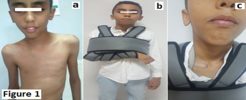
Case report: We report the case of a 12-year-old boy who presented with a right posterior SCD after a trauma occurring 24 h earlier at a sports activity. Clinical examination and radiological investigations by X-ray and computed tomography scan leads to diagnosis. He underwent an orthopedic closed reduction treatment without complications at 24 months of follow-up.
Conclusion: Management of posterior sternoclavicular dislocation is different between teams, with a tendency to use orthopedic reduction in case of fresh dislocation and absence of complications. Late diagnosis and management, as well as the presence of complications, may be difficult and life-threatening (due to vascular or tracheal compression).
Keywords: Sternoclavicular dislocation; child; orthopedic treatment; posterior displacement; sports.
Copyright: © Indian Orthopaedic Research Group.
Conflict of interest statement
Conflict of Interest: Nil
Figures


Similar articles
-
Traumatic posterior sternoclavicular joint dislocation in a child: a case report.Pan Afr Med J. 2014 Dec 17;19:386. doi: 10.11604/pamj.2014.19.386.5684. eCollection 2014. Pan Afr Med J. 2014. PMID: 25995782 Free PMC article.
-
Posterior Sternoclavicular Dislocation: A Case Report.J Educ Teach Emerg Med. 2021 Jan 15;6(1):V23-V25. doi: 10.21980/J8363Q. eCollection 2021 Jan. J Educ Teach Emerg Med. 2021. PMID: 37465538 Free PMC article.
-
Emergent Management of Traumatic Posterior Sternoclavicular Joint Dislocation: A Case Report and Literature Review.Cureus. 2021 Oct 23;13(10):e18996. doi: 10.7759/cureus.18996. eCollection 2021 Oct. Cureus. 2021. PMID: 34853739 Free PMC article.
-
Return to Sports After Closed Reduction of Acute Traumatic Posterior Sternoclavicular Joint Dislocations: A Systematic Review.Am J Sports Med. 2023 Sep;51(11):3076-3083. doi: 10.1177/03635465221131900. Epub 2022 Dec 6. Am J Sports Med. 2023. PMID: 36472354
-
Evaluation and Management of Sternoclavicular Dislocation in the Emergency Department.J Emerg Med. 2021 Nov;61(5):499-506. doi: 10.1016/j.jemermed.2021.07.038. Epub 2021 Sep 10. J Emerg Med. 2021. PMID: 34511297 Review.
References
-
- Gil-Albarova J, Rebollo-González S, Gómez-Palacio VE, Herrera A. Management of sternoclavicular dislocation in young children:Considerations about diagnosis and treatment of four cases. Musculoskelet Surg. 2013;97:137–43. - PubMed
-
- Garg S, Alshameeri ZA, Wallace WA. Posterior sternoclavicular joint dislocation in a child:A case report with review of literature. J Shoulder Elbow Surg. 2012;21:e11–6. - PubMed
-
- Honeycutt MW, Cox K, Michaeli D, Hulon B, Brewer J. Pediatric posterior sternoclavicular dislocation closed reduction and management. J Orthop Trauma. 2021;35:S11–2. - PubMed
-
- M Rousset P, Moreel S. Descamps, posterior sternoclavicular dislocation, Journal de Traumatologie du Sport. 2010;27((1)):14–19.
Publication types
LinkOut - more resources
Full Text Sources