

Impact of intrafraction motion in pancreatic cancer treatments with MR-guided adaptive radiation therapy
- PMID: 38162503
- PMCID: PMC10756668
- DOI: 10.3389/fonc.2023.1298099
Impact of intrafraction motion in pancreatic cancer treatments with MR-guided adaptive radiation therapy
Abstract
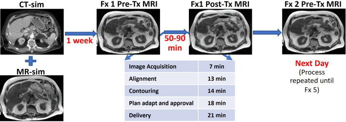
Purpose: The total time of radiation treatment delivery for pancreatic cancer patients with daily online adaptive radiation therapy (ART) on an MR-Linac can range from 50 to 90 min. During this period, the target and normal tissues undergo changes due to respiration and physiologic organ motion. We evaluated the dosimetric impact of the intrafraction physiological organ changes.
Methods: Ten locally advanced pancreatic cancer patients were treated with 50 Gy in five fractions with intensity-modulated respiratory-gated radiation therapy on a 0.35-T MR-Linac. Patients received both pre- and post-treatment volumetric MRIs for each fraction. Gastrointestinal organs at risk (GI-OARs) were delineated on the pre-treatment MRI during the online ART process and retrospectively on the post-treatment MRI. The treated dose distribution for each adaptive plan was assessed on the post-treatment anatomy. Prescribed dose volume histogram metrics for the scheduled plan on the pre-treatment anatomy, the adapted plan on the pre-treatment anatomy, and the adapted plan on post-treatment anatomy were compared to the OAR-defined criteria for adaptation: the volume of the GI-OAR receiving greater than 33 Gy (V33Gy) should be ≤1 cubic centimeter.
Results: Across the 50 adapted plans for the 10 patients studied, 70% were adapted to meet the duodenum constraint, 74% for the stomach, 12% for the colon, and 48% for the small bowel. Owing to intrafraction organ motion, at the time of post-treatment imaging, the adaptive criteria were exceeded for the duodenum in 62% of fractions, the stomach in 36%, the colon in 10%, and the small bowel in 48%. Compared to the scheduled plan, the post-treatment plans showed a decrease in the V33Gy, demonstrating the benefit of plan adaptation for 66% of the fractions for the duodenum, 95% for the stomach, 100% for the colon, and 79% for the small bowel.
Conclusion: Post-treatment images demonstrated that over the course of the adaptive plan generation and delivery, the GI-OARs moved from their isotoxic low-dose region and nearer to the dose-escalated high-dose region, exceeding dose-volume constraints. Intrafraction motion can have a significant dosimetric impact; therefore, measures to mitigate this motion are needed. Despite consistent intrafraction motion, plan adaptation still provides a dosimetric benefit.
Keywords: MR-guided radiation therapy; MR-linac; gastrointestinal motion; intrafraction motion; online adaptive radiation therapy; pancreatic cancer; respiratory-gated radiation therapy; stereotactic body radiation therapy (SBRT).
Copyright © 2023 Rusu, Cunningham, Arch, Chetty, Parikh and Dolan.
Conflict of interest statement
JC and JA report travel expenses from ViewRay outside of this work. PP and JD report grants, honoraria and travel expenses from ViewRay outside of this work. PP has clinical research funding from Speakers Board, ViewRay and ownership interest in Nuvaira, neither are related to this work. The remaining authors declare that the research was conducted in the absence of any commercial or financial relationships that could be construed as a potential conflict of interest.
Figures


Similar articles
-
Role of Daily Plan Adaptation in MR-Guided Stereotactic Ablative Radiation Therapy for Adrenal Metastases.Int J Radiat Oncol Biol Phys. 2018 Oct 1;102(2):426-433. doi: 10.1016/j.ijrobp.2018.06.002. Epub 2018 Jun 11. Int J Radiat Oncol Biol Phys. 2018. PMID: 29902559
-
Feasibility of ablative stereotactic body radiation therapy of pancreas cancer patients on a 1.5 Tesla magnetic resonance-linac system using abdominal compression.Phys Imaging Radiat Oncol. 2021 Jul 12;19:53-59. doi: 10.1016/j.phro.2021.07.006. eCollection 2021 Jul. Phys Imaging Radiat Oncol. 2021. PMID: 34307919 Free PMC article.
-
Investigation of Isotoxic Dose Escalation and Plan Quality with TDABC Analysis on a 0.35 T MR-Linac (MRL) System in Ablative 5-Fraction Stereotactic Magnetic Resonance-Guided Radiation Therapy (MRgRT) for Primary Pancreatic Cancer.J Clin Med. 2022 May 5;11(9):2584. doi: 10.3390/jcm11092584. J Clin Med. 2022. PMID: 35566712 Free PMC article.
-
Dose accumulation for MR-guided adaptive radiotherapy: From practical considerations to state-of-the-art clinical implementation.Front Oncol. 2023 Jan 26;12:1086258. doi: 10.3389/fonc.2022.1086258. eCollection 2022. Front Oncol. 2023. PMID: 36776378 Free PMC article. Review.
-
Adaptive Radiation Therapy in the Treatment of Lung Cancer: An Overview of the Current State of the Field.Front Oncol. 2021 Nov 29;11:770382. doi: 10.3389/fonc.2021.770382. eCollection 2021. Front Oncol. 2021. PMID: 34912715 Free PMC article. Review.
Cited by
-
Intrafractional Motion in Online-Adaptive Magnetic Resonance-Guided Radiotherapy of Adrenal Metastases Leads to Reduced Target Volume Coverage and Elevated Organ-at-Risk Doses.Cancers (Basel). 2025 Apr 30;17(9):1533. doi: 10.3390/cancers17091533. Cancers (Basel). 2025. PMID: 40361458 Free PMC article.
References
LinkOut - more resources
Full Text Sources