. 2023 Dec 14:14:1293828.
doi: 10.3389/fimmu.2023.1293828. eCollection 2023.

Soluble CD83 modulates human-monocyte-derived macrophages toward alternative phenotype, function, and metabolism

Affiliations

Soluble CD83 modulates human-monocyte-derived macrophages toward alternative phenotype, function, and metabolism

Katrin Peckert-Maier et al. Front Immunol. .

Abstract

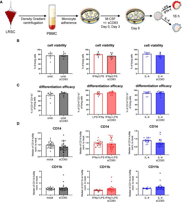
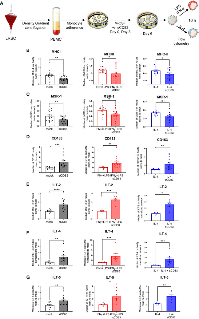
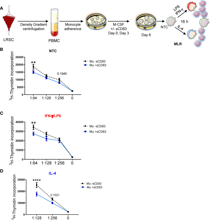
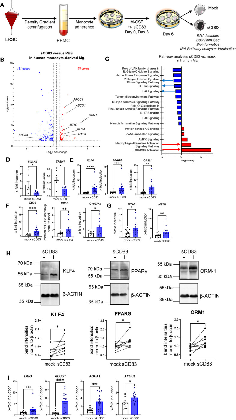
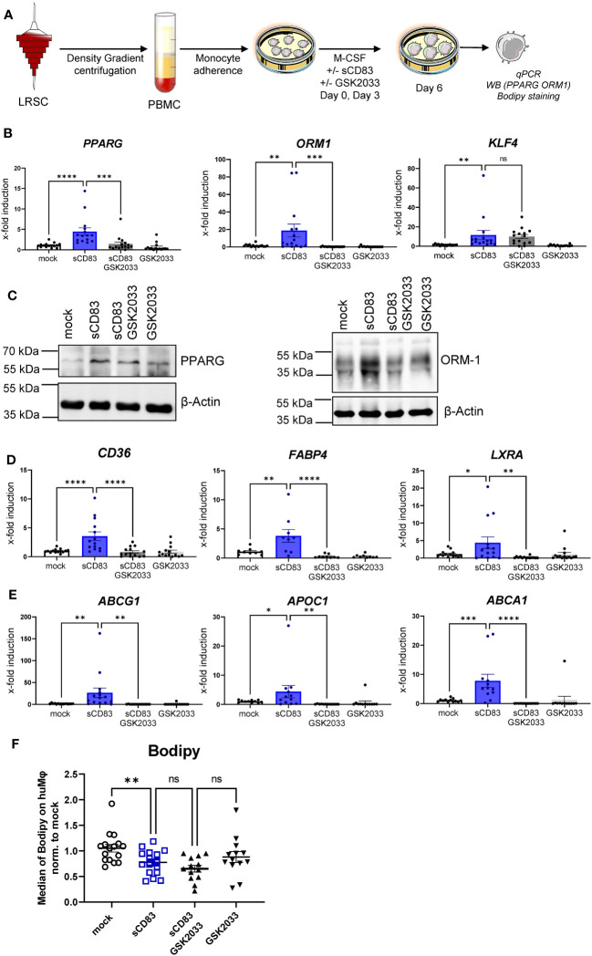
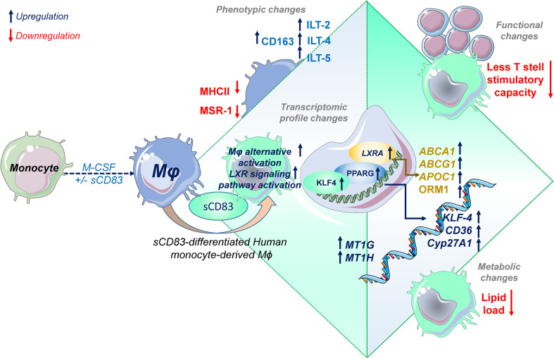
Alterations in macrophage (Mφ) polarization, function, and metabolic signature can foster development of chronic diseases, such as autoimmunity or fibrotic tissue remodeling. Thus, identification of novel therapeutic agents that modulate human Mφ biology is crucial for treatment of such conditions. Herein, we demonstrate that the soluble CD83 (sCD83) protein induces pro-resolving features in human monocyte-derived Mφ biology. We show that sCD83 strikingly increases the expression of inhibitory molecules including ILT-2 (immunoglobulin-like transcript 2), ILT-4, ILT-5, and CD163, whereas activation markers, such as MHC-II and MSR-1, were significantly downregulated. This goes along with a decreased capacity to stimulate alloreactive T cells in mixed lymphocyte reaction (MLR) assays. Bulk RNA sequencing and pathway analyses revealed that sCD83 downregulates pathways associated with pro-inflammatory, classically activated Mφ (CAM) differentiation including HIF-1A, IL-6, and cytokine storm, whereas pathways related to alternative Mφ activation and liver X receptor were significantly induced. By using the LXR pathway antagonist GSK2033, we show that transcription of specific genes (e.g., PPARG, ABCA1, ABCG1, CD36) induced by sCD83 is dependent on LXR activation. In summary, we herein reveal for the first time mechanistic insights into the modulation of human Mφ biology by sCD83, which is a further crucial preclinical study for the establishment of sCD83 as a new therapeutical agent to treat inflammatory conditions.

Keywords: LXR pathway; alternative activation; checkpoint molecule; human-monocyte-derived macrophages; soluble CD83.


Conflict of interest statement

The authors declare that the research was conducted in the absence of any commercial or financial relationships that could be construed as a potential conflict of interest.

Figures

Figure 1
Figure 1
Soluble CD83 does not interfere with viability nor differentiation efficacy of human Mφ. (A) Experimental set up to check whether sCD83 affects cell viability or differentiation efficacy of human monocyte-derived Mφ. PBMCs were isolated via density gradient centrifugation and subsequently monocytes were seeded for adherence. Mφ were differentiated from monocytes in the presence of M-CSF (20 ng/ml) and sCD83 (25 µg/ml) or the corresponding amount of DPBS was added on day 0 as well as day 3 during the differentiation process. Mφ were subsequently seeded and polarized via LPS (100 ng/ml) +IFN-γ (300 U/ml), or IL-4 (20 ng/ml). (B) Cell viability assessment of human monocyte-derived Mφ generated +/-sCD83 by flow cytometry. (C) sCD83 has no influence on differentiation efficacy of human monocyte-derived Mφ (D) Assessment of expression levels of CD14 (upper row) as well as CD11b (lower row) are not influenced by sCD83 when present during Mφ differentiation. Statistical analyses were performed by One-way ANOVA or the appropriate corresponding non-parametric test. Data are represented as mean ± SEM. Experiments were performed at least three times. One dot per bar graph represent one donor. The absence of asterisks indicates that there is no statistical significance.
Figure 2
Figure 2
Soluble CD83 strikingly modulates the phenotype of human monocyte-derived Mφ. (A) Experimental setup for the analyses of the phenotype of Mφ differentiated in the presence of sCD83 or PBS (referred to as mock). PBMCs were isolated via density gradient centrifugation, and subsequently, monocytes were seeded for adherence. Monocytes were differentiated into Mφ in the presence of M-CSF (20 ng/ml), +/− sCD83 (25 µg/ml). sCD83 was added on day 0 as well as day 3 during the differentiation process. Mφ were subsequently seeded and polarized via LPS (100 ng/ml) +IFN-γ (300 U/ml), or IL-4 (20 ng/ml) for 16 h. Subsequently, human monocyte-derived Mφ were analyzed by flow cytometry for the expression of (B) MHC-II, (C) MSR-1, (D) CD163, (E) ILT-2, (F) ILT-4, and (G) ILT-5. Data are represented as mean ± SEM. Statistical analysis was performed using a Mann–Whitney U test. Experiments were performed at least three times. One dot per bar graph represents one donor. n.s., not significant, which indicates there is no statistical significance; *p< 0.05; **< 0.01; *** p< 0.001; **** p< 0.0001.
Figure 3
Figure 3
Differentiation of human monocyte-derived Mφ in the presence of sCD83 results in less T cell stimulatory capacity. (A) Experimental set up for the analyses of the allogeneic T cell stimulatory capacity of human Mφ differentiated in the presence of sCD83 or PBS (referred to as mock). PBMCs were isolated via density gradient centrifugation and subsequently monocytes were seeded for adherence. Monocytes were differentiated into Mφ in the presence of M-CSF (20 ng/ml), +/- sCD83 (25 µg/ml). sCD83 was added on day 0 as well as day 3 during the differentiation process. Mφ were subsequently seeded and polarized LPS (100 ng/ml) +IFN-γ (300 U/ml), or IL-4 (20 ng/ml). Subsequently, human monocyte-derived Mφ were analyzed for the capacity to stimulate alloreactive T cells using MLR assays. (A–D) Human monocyte-derived Mφ Untreated (B), LPS+IFNγ (C) IL-4 (D) stimulated sCD83-differentiated human Mφ show decreased capacity to stimulate alloreactive T cells regardless of the preceding stimulation compared to control Mφ N ≥ 7; Experiments were performed at least 4 times for each stimulation. Data are represented as mean ± SEM. Statistical analysis was performed using a Two-way ANOVA or the appropriate corresponding non-parametric test. Experiments were performed at least three times. One dot per bar graph represent one donor n.s., not significant, which indicates there is no statistical significance; ** < 0.01; **** p < 0.0001.
Figure 4
Figure 4
Soluble CD83-differentiated human monocyte-derived Mφ show distinct profile changes toward alternative activation on mRNA as well as on the protein level. (A) Experimental setup for the bulk RNA sequencing experiment and verification on mRNA as well as on the protein level by qPCR and Western blot. PBMCs were isolated via density gradient centrifugation, and subsequently, monocytes were seeded for adherence. Monocytes were differentiated into Mφ in the presence of M-CSF (20 ng/ml) and +/− sCD83 (25 µg/ml). sCD83 was added on day 0 as well as day 3 during the differentiation process. Subsequently, Mφ were harvested on day 6 of differentiation and RNA sequencing analyses as well as verification on protein as well as mRNA levels were performed by Western blotting as well as qPCR. (B) Volcano plot of RNA sequencing analyses of human monocyte-derived Mφ differentiated in the presence of sCD83 versus mock-treated Mφ (n = 3). On the right-hand side of the logFold change 0 value, significantly upregulated gene transcripts (red) are displayed, whereas the dots on the left-hand side represent significantly downregulated transcripts (blue) with logFC ≥0.585, respectively. On the Y-axis, the p-value is displayed. (C) Transcriptomic data were analyzed using the Ingenuity Pathway Analysis (IPA) software from QIAGEN’s revealing downregulation of inflammatory pathways and enhanced upregulation of resolving Mφ pathways including alternative activation as well as LXR activation. (D) qPCR analyses for mRNA expression analyses of HIF-1A target genes EGLN3 and TREM-1, which are associated with CAM polarization in +/−sCD83 differentiated Mφ. (E) qPCR analyses for mRNA expression analyses of genes, such as KLF-4, PPARγ, and ORM1, which are associated with AAM in +/−sCD83-differentiated Mφ. (F) qPCR/flow cytometric analyses for PPARγ target genes, such as CD36, CD36, and Cyp27A1, which are associated with AAM in +/−sCD83-differentiated Mφ. (G) qPCR analyses for mRNA expression analyses for MT1G as well as MT1H, which are associated with anti-inflammatory properties. (H) Western blot analyses of whole-cell lysates of human Mφ-differentiated +/− sCD83 for assessment of KLF-4, PPARG, and ORM-1 protein levels. Quantification of Western blots was performed using the ImageJ software and β-ACTIN served as loading control. (I) qPCR analyses for mRNA expression analyses for LXRa (NR1H3) and its target genes, such as ABCG1, ABCA1, and APOC1, which are associated with AAM in +/− sCD83-differentiated Mφ. Data are represented as mean ± SEM. Statistical analysis was performed using a one-way ANOVA or the appropriate corresponding non-parametric test. Experiments were performed at least three times. One dot per bar graph represents one donor. n.s., not significant, which indicates there is no statistical significance; *p< 0.05; **< 0.01; ***p< 0.001; ****p< 0.0001.
Figure 5
Figure 5
Soluble CD83-induced transcriptional changes are partly dependent on LXR pathway activation. (A) Experimental setup for the LXR pathway blocking experiments. PBMCs were isolated via density gradient centrifugation, and subsequently, monocytes were seeded for adherence. Monocytes were differentiated into Mφ in the presence of M-CSF (20 ng/ml) and +/− sCD83 (25 µg/ml). sCD83 was added on day 0 as well as day 3 during the differentiation process. GSK2033 (1 mM) was applied 1 h before sCD83 administration. Subsequently, cells were harvested for subsequent analyses. (B) qPCR analyses for mRNA expression of PPARG (first bar graph), ORM-1 (second bar graph), and KLF-4 (third bar graph) of mock-, sCD83-, sCD83 + GSK2033-, or GSK2033-treated Mφ. (C) Western blot analyses of whole-cell lysates of human monocyte-derived Mφ differentiated +/− sCD83 for assessment of PPARG as well as ORM-1 protein levels. (D) qPCR analyses for mRNA expression of CD36 (first bar graph), FABP4 (second bar graph), LXRa (third bar graph) of mock-, sCD83-, sCD83 + GSK2033-, or GSK2033-treated Mφ. (E) qPCR analyses for mRNA expression of ABCG1 (first bar graph), APOC1 (second bar graph), ABCA1 (third bar graph) of mock-, sCD83-, sCD83 + GSK2033-, or GSK2033-treated Mφ. (F) Flow cytometric analyses using Bodipy staining, which is a fluorescent dye that stains lipid droplets in human monocyte-derived Mφ. Data are represented as mean ± SEM. Statistical analysis was performed using a one-way ANOVA or the appropriate corresponding non-parametric test. Experiments were performed at least three times. One dot per bar graph represents one donor. n.s., not significant, which indicates there is no statistical significance; *p< 0.05; **< 0.01; *** p< 0.001; **** p< 0.0001.
Figure 6
Figure 6
Soluble CD83 induces phenotypical, functional, and metabolic changes in human monocyte-derived Mφ. Administration of sCD83 to Mφ differentiation results in upregulation of pro-resolving receptors (CD163, ILT-2, ILT-4, ILT-5), whereas activation markers including MSR-1 or MHC-II are downregulated, which results in less T-cell stimulatory capacity of sCD83-differentiated Mφ. Administration of sCD83 to Mφ differentiation results in profound changes in transcriptome, including induction of pathways associated with alternative activation as well as liver X receptor pathway activation in Mφ. To this end, Mφ treated with sCD83 show increased expression of transcription factors KLF4, PPARG, and LXRA, which are characteristic for human alternatively activated Mφ. In line with that, sCD83-treated Mφ show enhanced levels of well-characterized target genes, such as CD36, KLF-4, CYP27A1, ABCA1, ABCG1, and ORM1, indicating modulation of the lipid metabolic state of sCD83-differentiated Mφ, which results in less lipid load. In summary, we present for the first time data on modulation of human Mφ by sCD83, which represents a further preclinical step for the development of sCD83 as a new therapeutic agent for the treatment of inflammatory conditions.

Similar articles

Cited by

References

    1. Xue J, Schmidt SV, Sander J, Draffehn A, Krebs W, Quester I, et al. . Transcriptome-based network analysis reveals a spectrum model of human macrophage activation. Immunity (2014) 40(2):274–88. doi: 10.1016/j.immuni.2014.01.006 - DOI - PMC - PubMed
    1. Viola A, Munari F, Sanchez-Rodriguez R, Scolaro T, Castegna A. The metabolic signature of macrophage responses. Front Immunol (2019) 10:1462. doi: 10.3389/fimmu.2019.01462 - DOI - PMC - PubMed
    1. Eming SA, Murray PJ, Pearce EJ. Metabolic orchestration of the wound healing response. Cell Metab (2021) 33(9):1726–43. doi: 10.1016/j.cmet.2021.07.017 - DOI - PubMed
    1. Martinez FO, Gordon S. The M1 and M2 paradigm of macrophage activation: time for reassessment. F1000Prime Rep (2014) 6:13. doi: 10.12703/P6-13 - DOI - PMC - PubMed
    1. de Groot AE, Pienta KJ. Epigenetic control of macrophage polarization: implications for targeting tumor-associated macrophages. Oncotarget (2018) 9(29):20908–27. doi: 10.18632/oncotarget.24556 - DOI - PMC - PubMed

Publication types