

Age estimation from mandibles in Malay: A 2D geometric morphometric analysis
- PMID: 38162871
- PMCID: PMC10757314
- DOI: 10.1016/j.jtumed.2023.05.020
Age estimation from mandibles in Malay: A 2D geometric morphometric analysis
Abstract
Objectives: In this study, the sizes and forms of mandibles in various age groups of the Malay population were measured and compared.
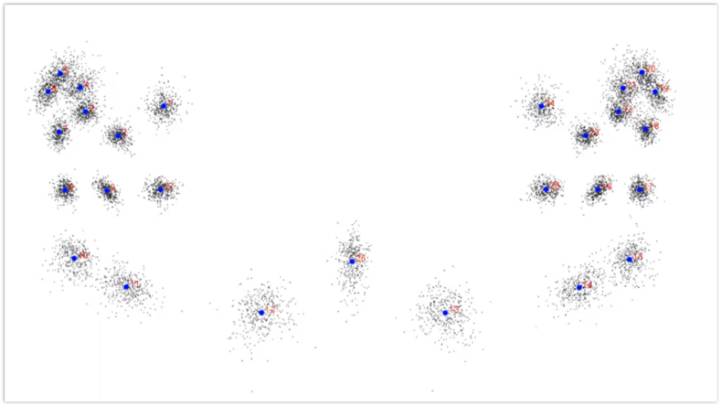
Methods: Geometric morphometric (GM) analysis of mandibles from 400 dental panoramic tomography (DPT) specimens was conducted. The MorphoJ program was used to perform generalized Procrustes analysis (GPA), Procrustes ANOVA, principal component analysis (PCA), discriminant function analysis (DFA), and canonical variate analysis (CVA). In the tpsDig2 program, the 27 landmarks were applied to the DPT radiographs. Variations in mandibular size and form were categorized into four age groups: group 1 (15-24 years), group 2 (25-34 years), group 3 (35-44 years), and group 4 (45-54 years).
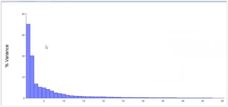
Results: The diversity in mandibular shape among the first eight principal components was 81%. Procrustes ANOVA revealed significant shape differences (P < 0.001) among age groups. Mahalanobis distances indicated substantial differences among all age groups; group 1 and group 4 scored highest, at 2.114. The ranges for the cross-validation and discriminant function tests were 90-72% and 81-49%, respectively.
Conclusion: GM analysis through radiography is a simple, non-invasive, and non-destructive method of estimating age by using the mandible. GM analysis is unique because it can visualize the changes in mandible shape among age groups. This method should aid in age identification in forensic odontology investigations.
أهداف البحث: تهدف هذه الدراسة إلى قياس ومقارنة حجم وشكل الفك السفلي في مجموعات عمرية مختلفة من السكان الماليزيين.
طرق البحث: أجري تحليل المورفومتريا الهندسية للفك السفلي باستخدام 400 عينة من التصوير المقطعي المقلاعي السني. استخدم برنامج مورفوجي لإجراء تحليل بروكرست العام، وتحليل تباين بروكرست، وتحليل المكونات الرئيسية، وتحليل الدالة التمييزية، وتحليل المتغيرات الكنسية. تم تطبيق ما مجموعه 27 علامة بارزة على صور التصوير المقطعي المقلاعي السني باستخدام برنامج "ت ب اس د آي ج 2". لتصنيف التغيرات في حجم وشكل الفك السفلي، تم إنشاء أربع مجموعات عمرية: المجموعة 1 (15-24 عاما)، والمجموعة 2 (25-34 عاما)، والمجموعة 3 (35-44 عاما)، والمجموعة 4 (45-54 عاما).
النتائج: أظهرت التغييرات في أشكال الفك السفلي متغيرا بنسبة 81% على أساس المكونات الرئيسية الثمانية الأولى. كشف تحليل تباين بروكرست عن اختلافات كبيرة في الشكل بين المجموعات العمرية. أظهرت مسافات ماهالانوبيس اختلافات كبيرة بين جميع المجموعات العمرية، حيث سجلت المجموعة 1 والمجموعة 4 أعلى قيمة بـ 2.114. تراوحت نسب الصحة لاختبارات التحقق المتقاطع بين 90% و72% ولاختبارات الدالة التمييزية بين 81% و49%.
الاستنتاجات: في الختام، يعتبر استخدام الأشعة السينية لتحليل المورفومتريا الهندسية طريقة بسيطة وغير ضارة وغير تدميرية لتقدير العمر باستخدام الفك السفلي. يعتبر تحليل المورفومتريا الهندسية فريدا لأنه يمكن تصوير تغييرات شكل الفك السفلي بين المجموعات العمرية المختلفة. تعتبر هذه الطريقة مفيدة لتحديد العمر في التحقيقات الطبية الشرعية في طب الأسنان.
Keywords: Age estimation; Dental panoramic tomography; Geometric morphometric; Identification; Mandible.
© 2023 The Authors.
Figures


Similar articles
-
Geometric morphometric evaluation of mandibles of four sheep breeds: Bardoka, İvesi, Polish Mountain sheep and Turcana.Anat Histol Embryol. 2024 May;53(3):e13048. doi: 10.1111/ahe.13048. Anat Histol Embryol. 2024. PMID: 38706190
-
A geometric morphometric evaluation of facial hard tissue patterns.J Orthod Sci. 2022 May 4;11:24. doi: 10.4103/jos.jos_199_21. eCollection 2022. J Orthod Sci. 2022. PMID: 35754421 Free PMC article.
-
Evaluation of sex dimorphism of the mandible with geometric morphometric analysis: conventional and reconstructed panoramic radiography study.Surg Radiol Anat. 2023 Nov;45(11):1497-1504. doi: 10.1007/s00276-023-03201-z. Epub 2023 Jul 17. Surg Radiol Anat. 2023. PMID: 37460704
-
[Principles and methods of geometric morphometrics].Zh Obshch Biol. 2002 Nov-Dec;63(6):473-93. Zh Obshch Biol. 2002. PMID: 12510587 Review. Russian.
-
The use of the geometric morphometric method to illustrate shape difference in the skulls of different-aged horses.Vet Res Commun. 2020 Nov;44(3-4):137-145. doi: 10.1007/s11259-020-09779-8. Epub 2020 Jul 23. Vet Res Commun. 2020. PMID: 32700122 Free PMC article. Review.
References
-
- Adams D.C., Rohlf F.J., Slice D.E. A field comes of age: geometric morphometrics in the 21st century. Hystrix. 2013;24(1) doi: 10.4404/hystrix-24.1-6283. - DOI
LinkOut - more resources
Full Text Sources
