

Microsurgical partial trapping for the treatment of unclippable vertebral artery aneurysms: Experience from 27 patients and review of literature
- PMID: 38163051
- PMCID: PMC10755825
- DOI: 10.1016/j.wnsx.2023.100256
Microsurgical partial trapping for the treatment of unclippable vertebral artery aneurysms: Experience from 27 patients and review of literature
Abstract
Background: The efficacy and safety of partial trapping for the treatment of unclippable vertebral artery aneurysms (UVAs) are still questionable. The partial trapping method (proximal or distal occlusion) was used in the treatment of aneurysms to simplify the surgical procedure and avoid postoperative complications.
Methods: This study included 27 patients with UVAs who underwent microsurgical partial trapping between January 2015 and August 2022, and their postoperative outcomes and complications were retrospectively reviewed and evaluated.
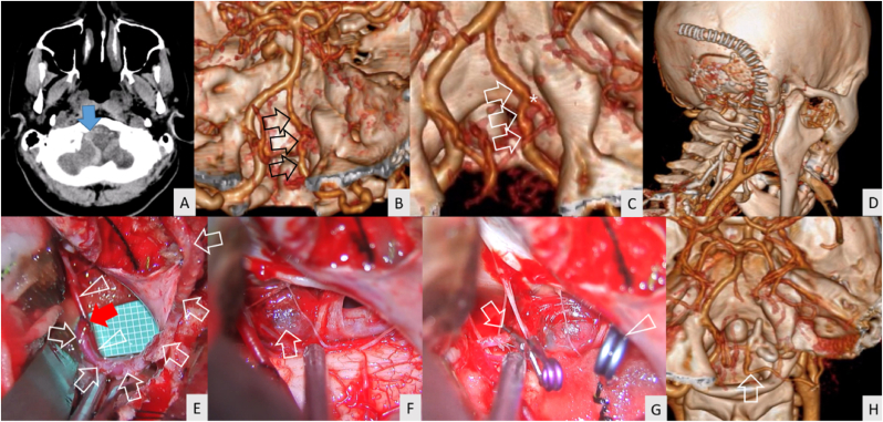
Results: Ruptured UVAs were detected in 25 (92.6%) patients, and 13 (48.1%) patients had poor-grade status. Fusiform dissection, dissecting, and fusiform aneurysms were observed in 17 (63%), 7 (25.9%), and 3 (11.1%) patients, respectively. By location, preposterior inferior cerebellar artery (PICA), PICA, post- PICA, and non-PICA types were noted in 7 (25.9%), 9 (33.3%), 6 (22.2%), and 5 (18.5%) patients, respectively. Microsurgical partial trapping was performed in all patients (blind-alley formation in 96.3%). Complete aneurysm obliteration was achieved in 26 (96.3%) patients. Immediate complete obliteration was achieved in 21 (77.8%) patients, delayed thrombosis within 7 days in 5 (18.5%), and nearly complete obliteration in 1 (3.7%). No re-bleeding was detected in all patients. Favorable outcomes 3 months after the operation were achieved by 92.9% of the patients in the good-grade group and 85.2% overall.
Conclusions: Microsurgical partial trapping, especially the blind-alley formation technique, was a safe and effective treatment of UVAs with high rates of aneurysm thrombosis. The appropriate sites for clip occlusion were dependent on the angioarchitecture of UVAs.
Keywords: Blind-alley formation; Incomplete trapping; Microsurgery; Partial trapping; Unclippable aneurysm; Vertebral artery aneurysm.
© 2023 The Authors.
Conflict of interest statement
The authors declare that they have no known competing financial interests or personal relationships that could have appeared to influence the work reported in this paper.
Figures


Similar articles
-
Blind-Alley Formation and Occipital Artery-Posterior Inferior Cerebellar Artery Bypass for the Treatment of Unclippable Vertebral Artery Aneurysms with Posterior Inferior Cerebellar Artery Involvement.World Neurosurg. 2020 Jun;138:e539-e550. doi: 10.1016/j.wneu.2020.02.174. Epub 2020 Mar 7. World Neurosurg. 2020. PMID: 32156594
-
Microsurgical Treatment of Vertebral Artery Dissection: Surgical Strategies and Treatment Outcomes.World Neurosurg. 2022 Mar;159:e375-e388. doi: 10.1016/j.wneu.2021.12.057. Epub 2021 Dec 22. World Neurosurg. 2022. PMID: 34954059
-
Vertebrobasilar dissecting aneurysms: microsurgical management in 42 patients.J Neurosurg. 2021 Dec 10;137(2):393-401. doi: 10.3171/2021.9.JNS21397. Print 2022 Aug 1. J Neurosurg. 2021. PMID: 34891141
-
Endovascular parent vessel sacrifice in ruptured dissecting vertebral and posterior inferior cerebellar artery aneurysms: clinical outcomes and review of the literature.J Neurointerv Surg. 2016 Aug;8(8):796-801. doi: 10.1136/neurintsurg-2015-011732. Epub 2015 Aug 3. J Neurointerv Surg. 2016. PMID: 27417905 Review.
-
Subarachnoid hemorrhage from vertebral artery dissecting aneurysms involving the origin of the posteroinferior cerebellar artery: report of two cases and review of the literature.Neurosurgery. 2000 Jan;46(1):196-200; discussion 200-1. Neurosurgery. 2000. PMID: 10626950 Review.
References
-
- Iihara K., Sakai N., Murao K., et al. Dissecting aneurysms of the vertebral artery: a management strategy. J Neurosurg. 2002;97:259–267. - PubMed
-
- Spetzler R.F., McDougall C.G., Zabramski J.M., et al. The barrow ruptured aneurysm trial: 6-year results. J Neurosurg. 2015;123:609–617. - PubMed
-
- Wallace A.N., CreveCoeur T.S., Grossberg J.A., et al. Impact of aneurysm morphology on safety and effectiveness of flow diverter treatment of vertebrobasilar aneurysms. J Neuroradiol. 2019;46:401–410. - PubMed
-
- Aboukaïs R., Zairi F., Boustia F., Bourgeois P., Leclerc X., Lejeune J.P. Vertebral artery-posterior inferior cerebellar artery convergence aneurysms treated by endovascular or surgical treatment: mid- and long-term outcome. Neurochirurgie. 2016;62:72–77. - PubMed
LinkOut - more resources
Full Text Sources
