

Faecalibacterium prausnitzii promotes intestinal epithelial IL-18 production through activation of the HIF1α pathway
- PMID: 38163085
- PMCID: PMC10755969
- DOI: 10.3389/fmicb.2023.1298304
Faecalibacterium prausnitzii promotes intestinal epithelial IL-18 production through activation of the HIF1α pathway
Abstract
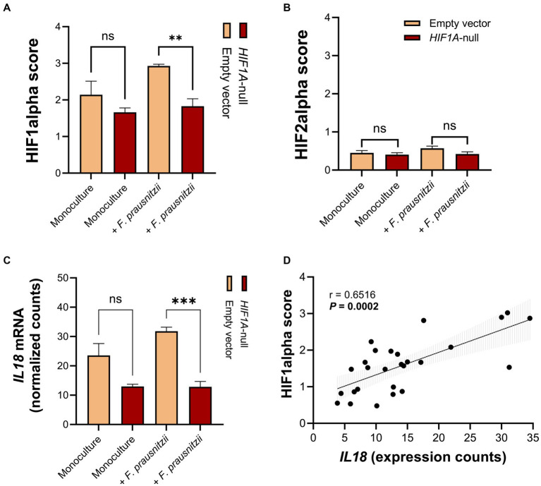
Introduction: Intestinal epithelial cells produce interleukin-18 (IL-18), a key factor in promoting epithelial barrier integrity. Here, we analyzed the potential role of gut bacteria and the hypoxia-inducible factor 1α (HIF1α) pathway in regulating mucosal IL18 expression in inflammatory bowel disease (IBD).
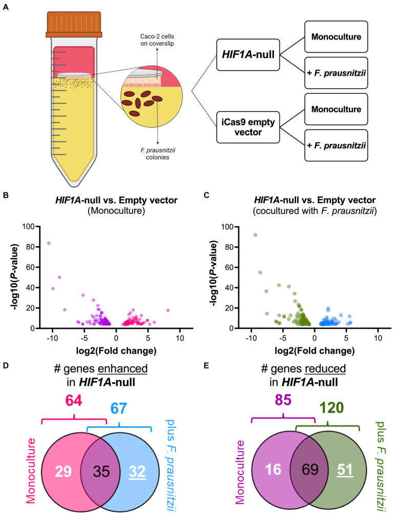
Methods: Mucosal samples from patients with IBD (n = 760) were analyzed for bacterial composition, IL18 levels and HIF1α pathway activation. Wild-type Caco-2 and CRISPR/Cas9-engineered Caco-2-HIF1A-null cells were cocultured with Faecalibacterium prausnitzii in a "Human oxygen-Bacteria anaerobic" in vitro system and analyzed by RNA sequencing.
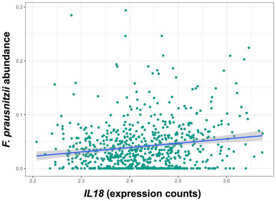
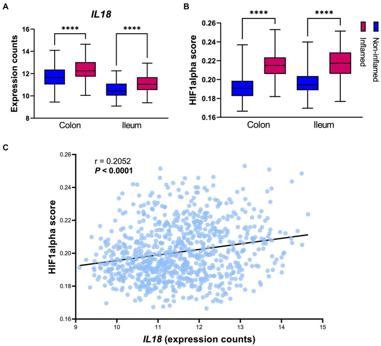
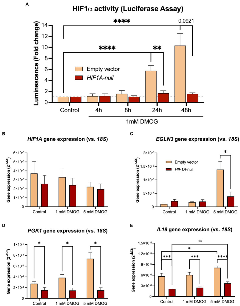
Results: Mucosal IL18 mRNA levels correlated positively with the abundance of mucosal-associated butyrate-producing bacteria, in particular F. prausnitzii, and with HIF1α pathway activation in patients with IBD. HIF1α-mediated expression of IL18, either by a pharmacological agonist (dimethyloxallyl glycine) or F. prausnitzii, was abrogated in Caco-2-HIF1A-null cells.
Conclusion: Butyrate-producing gut bacteria like F. prausnitzii regulate mucosal IL18 expression in a HIF1α-dependent manner that may aid in mucosal healing in IBD.
Keywords: Faecalibacterium; HIF1α; HoxBan in vitro coculture system; IBD; IL-18; epithelial-bacteria interaction; intestine.
Copyright © 2023 Fagundes, Bravo-Ruiseco, Hu, Kierans, Weersma, Taylor, Dijkstra, Harmsen and Faber.
Conflict of interest statement
GD received research grant from Royal DSM and speaker’s fees from Janssen Pharmaceuticals, Pfizer and Abbvie. RW acted as consultant for Takeda and received unrestricted grants from Takeda, Johnson & Johnson, Tramedico and Ferring, and received speaker’s fee from MSD, Abbvie and Janssen Pharmaceuticals. The remaining authors declare that the research was conducted in the absence of any commercial or financial relationships that could be construed as a potential conflict of interest.
Figures
Similar articles
-
HIF1α-Dependent Induction of TFRC by a Combination of Intestinal Inflammation and Systemic Iron Deficiency in Inflammatory Bowel Disease.Front Physiol. 2022 Jun 8;13:889091. doi: 10.3389/fphys.2022.889091. eCollection 2022. Front Physiol. 2022. PMID: 35755436 Free PMC article.
-
Live Faecalibacterium prausnitzii Does Not Enhance Epithelial Barrier Integrity in an Apical Anaerobic Co-Culture Model of the Large Intestine.Nutrients. 2017 Dec 12;9(12):1349. doi: 10.3390/nu9121349. Nutrients. 2017. PMID: 29231875 Free PMC article.
-
Inulin-grown Faecalibacterium prausnitzii cross-feeds fructose to the human intestinal epithelium.Gut Microbes. 2021 Jan-Dec;13(1):1993582. doi: 10.1080/19490976.2021.1993582. Gut Microbes. 2021. PMID: 34793284 Free PMC article.
-
Development, validation and implementation of an in vitro model for the study of metabolic and immune function in normal and inflamed human colonic epithelium.Dan Med J. 2015 Jan;62(1):B4973. Dan Med J. 2015. PMID: 25557335 Review.
-
Systematic review and meta-analysis of the role of Faecalibacterium prausnitzii alteration in inflammatory bowel disease.J Gastroenterol Hepatol. 2021 Feb;36(2):320-328. doi: 10.1111/jgh.15222. Epub 2020 Sep 7. J Gastroenterol Hepatol. 2021. PMID: 32815163
Cited by
-
Celastrol alleviates esophageal stricture in rats by inhibiting NLR family pyrin domain containing 3 activation.World J Gastroenterol. 2025 Jun 21;31(23):106949. doi: 10.3748/wjg.v31.i23.106949. World J Gastroenterol. 2025. PMID: 40575338 Free PMC article.
-
Crosstalk between hypoxia-inducible factor-1α and short-chain fatty acids in inflammatory bowel disease: key clues toward unraveling the mystery.Front Immunol. 2024 Mar 28;15:1385907. doi: 10.3389/fimmu.2024.1385907. eCollection 2024. Front Immunol. 2024. PMID: 38605960 Free PMC article. Review.
-
Bacterial extracellular vesicles at the interface of gut microbiota and immunity.Gut Microbes. 2024 Jan-Dec;16(1):2396494. doi: 10.1080/19490976.2024.2396494. Epub 2024 Sep 28. Gut Microbes. 2024. PMID: 39340209 Free PMC article. Review.
-
Biological characteristics, immune infiltration and drug prediction of PANoptosis related genes and possible regulatory mechanisms in inflammatory bowel disease.Sci Rep. 2025 Jan 15;15(1):2033. doi: 10.1038/s41598-024-84911-1. Sci Rep. 2025. PMID: 39814753 Free PMC article.
References
-
- Bao C. H., Wang C. Y., Li G. N., Yan Y. L., Wang D., Jin X. M., et al. . (2019). Effect of mild moxibustion on intestinal microbiota and NLRP6 inflammasome signaling in rats with post-inflammatory irritable bowel syndrome. World J. Gastroenterol. 25, 4696–4714. doi: 10.3748/wjg.v25.i32.4696 - DOI - PMC - PubMed
LinkOut - more resources
Full Text Sources
Molecular Biology Databases
Miscellaneous