

Prone extension views in cervical MRI: A case-driven Novel approach
- PMID: 38163148
- PMCID: PMC10755320
- DOI: 10.1016/j.heliyon.2023.e23251
Prone extension views in cervical MRI: A case-driven Novel approach
Abstract
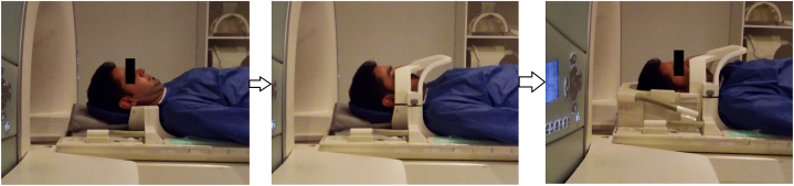
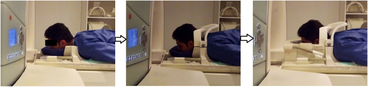
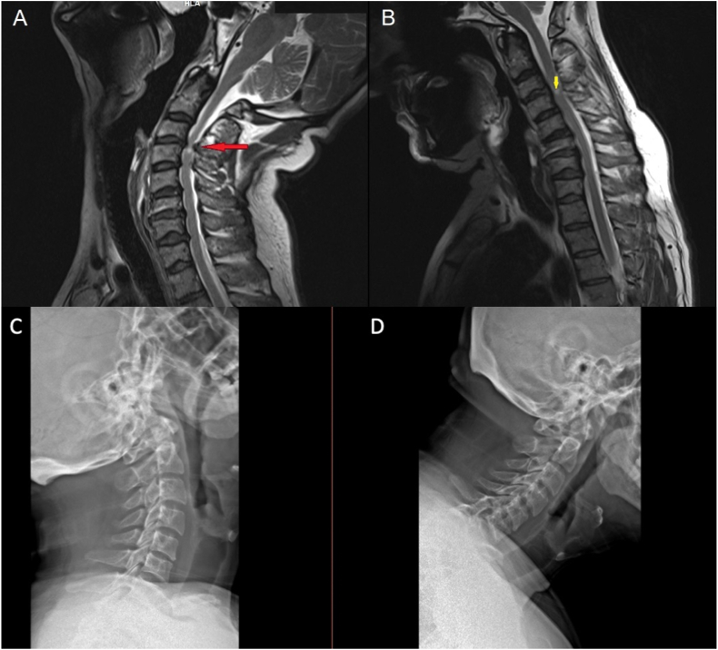
The cervical spinal canal has a wide range of motion and specific biomechanics involved with different pathologies that may cause dynamic cord compressions. This study has introduced new protocol for acquiring an extension view of cervical MRI to assess dynamic cervical spinal canal compromise. We posit that dynamic MRI comprising extension view in prone position could be a practical option when deciding the best approach in treating challenging patients.
Keywords: Cervical spine; Cord compression; Dynamic MRI; Prone position.
© 2023 The Authors.
Conflict of interest statement
The authors declare that they have no known competing financial interests or personal relationships that could have appeared to influence the work reported in this paper.
Figures
Similar articles
-
The value of dynamic MRI in cervical spondylotic myelopathy: About 24 cases.Ann Med Surg (Lond). 2022 Sep 22;83:104717. doi: 10.1016/j.amsu.2022.104717. eCollection 2022 Nov. Ann Med Surg (Lond). 2022. PMID: 36389194 Free PMC article.
-
Kinetic change of spinal cord compression on flexion-extension magnetic resonance imaging in cervical spine.Clin Neurol Neurosurg. 2018 Nov;174:86-91. doi: 10.1016/j.clineuro.2018.09.017. Epub 2018 Sep 10. Clin Neurol Neurosurg. 2018. PMID: 30219623
-
Dynamic Cord Compression Causing Cervical Myelopathy.Neurospine. 2019 Sep;16(3):448-453. doi: 10.14245/ns.1938020.101. Epub 2019 Jul 24. Neurospine. 2019. PMID: 31607076 Free PMC article.
-
[The dorsal approach in degeneratively changed cervical spine].Orthopade. 1996 Nov;25(6):533-41. doi: 10.1007/s001320050057. Orthopade. 1996. PMID: 8999408 Review. German.
-
Pediatric cervical kyphosis in the MRI era (1984-2008) with long-term follow up: literature review.Childs Nerv Syst. 2022 Feb;38(2):361-377. doi: 10.1007/s00381-021-05409-z. Epub 2021 Nov 22. Childs Nerv Syst. 2022. PMID: 34806157 Review.
References
-
- Shedid D., Benzel E.C. Cervical spondylosis anatomy: pathophysiology and biomechanics. Neurosurgery. 2007;60(suppl_1):S1–S7. - PubMed
-
- Jinkins J.R., Dworkin J.S., Green C.A., et al. Upright, weight-bearing, dynamic-kinetic magnetic resonance imaging of the spine-review of the first clinical results. JOURNAL-HONG KONG Coll Radiol. 2003;6:55–74.
-
- Zeitoun D., El Hajj F., Sariali E., et al. Evaluation of spinal cord compression and hyperintense intramedullary lesions on T2-weighted sequences in patients with cervical spondylotic myelopathy using flexion-extension MRI protocol. Spine J. 2015;15(4):668–674. - PubMed
-
- Burke C.J., Samim M., Alizai H., et al. Clinical feasibility of 2D dynamic sagittal HASTE flexion-extension imaging of the cervical spine for the assessment of spondylolisthesis and cervical cord impingement. Eur. J. Radiol. 2021;134 - PubMed
Publication types
LinkOut - more resources
Full Text Sources
