. 2023 Dec 3;10(1):e23233.
doi: 10.1016/j.heliyon.2023.e23233. eCollection 2024 Jan 15.

LncRNA SNHG8 upregulates MUC5B to induce idiopathic pulmonary fibrosis progression by targeting miR-4701-5p

Affiliations

LncRNA SNHG8 upregulates MUC5B to induce idiopathic pulmonary fibrosis progression by targeting miR-4701-5p

Xiaoping Zhang et al. Heliyon. .

Abstract

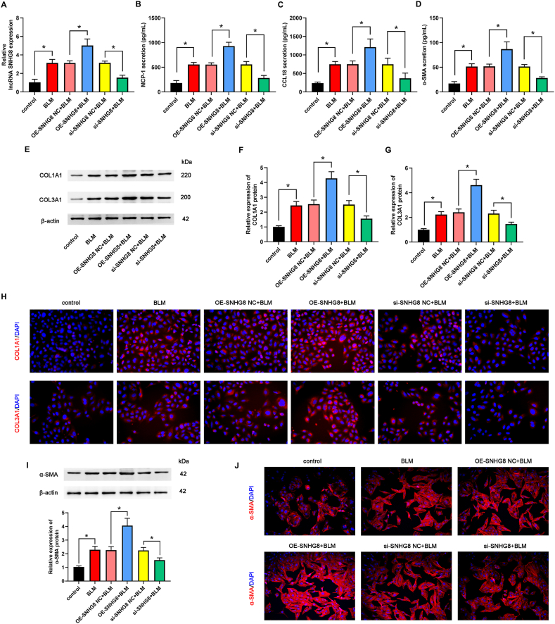
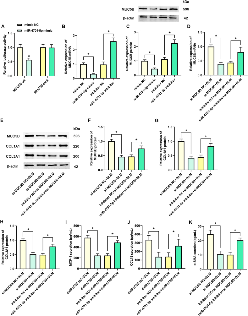
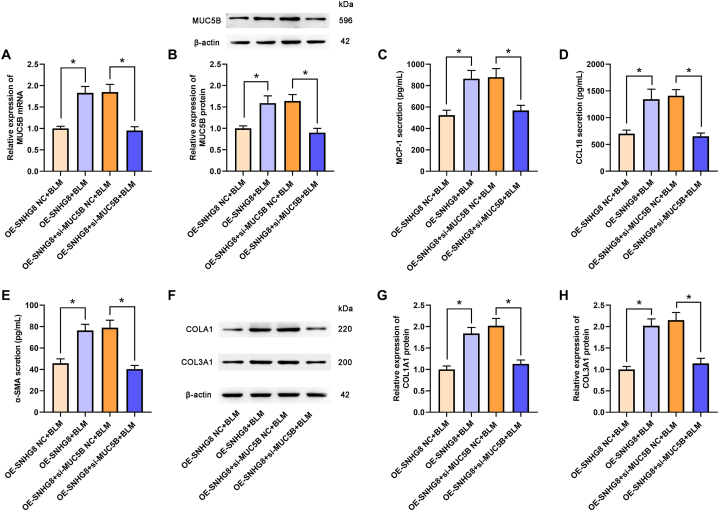
Long noncoding RNAs (lncRNAs) play a critical role in idiopathic pulmonary fibrosis (IPF); however, the underlying molecular mechanisms are unclear. Our study demonstrated that lncRNA small nucleolar RNA host gene 8 (SNHG8) was increased in bleomycin (BLM)-induced A549 cells. LncRNA SNHG8 overexpression further elevated fibrosis-related factors monocyte chemotactic protein 1 (MCP1), CC motif chemokine ligand 18 (CCL18), and α-smooth muscle actin (α-SMA), as well as increased collagen type I alpha-1 chain (COL1A1) and collagen type III alpha-1 chain (COL3A1). Meanwhile, lncRNA SNHG8 knockdown exhibited an opposite role in reducing BLM-induced pulmonary fibrosis. With regard to the mechanism, SNHG8 was then revealed to act as a competing endogenous RNA (ceRNA) for microRNA (miR)-4701-5p in regulating Mucin 5B (MUC5B) expression. Furthermore, the interactions between SNHG8 and miR-4701-5p, between miR-4701-5p and MUC5B, and between SNHG8 and MUC5B on the influence of fibrosis-related indicators were confirmed, respectively. In addition, SNHG8 overexpression enhanced the levels of transforming growth factor (TGF)-β1 and phosphorylation Smad2/3 (p-Smad2/3), which was suppressed by SNHG8 knockdown in BLM-induced A549 cells. Moreover, miR-4701-5p inhibitor-induced elevation of TGF-β1 and p-Smad2/3 was significantly suppressed by SNHG8 knockdown. In conclusion, SNHG8 knockdown attenuated pulmonary fibrosis progression by regulating miR-4701-5p/MUC5B axis, which might be associated with the modulation of TGF-β1/Smad2/3 signaling. These findings reveal that lncRNA SNHG8 may become a potential target for the treatment of IPF.

Keywords: IPF; MUC5B; TGF-β1/Smad2/3 signaling; lncRNA SNHG8; miR-4701-5p.


Conflict of interest statement

The authors declare that they have no known competing financial interests or personal relationships that could have appeared to influence the work reported in this paper.

Figures

Fig. 1
Fig. 1
SNHG8 and MUC5B expression were upregulated, and miR-4701-5p expression was downregulated in BLM-induced A549 cells. The expression of SNHG8 (A), miR-4701-5p (B), and MUC5B mRNA (C) were assessed by qPCR assay. (D) The protein expression of MUC5B was assessed by Western blot assay, and the relative quantitative analysis was shown. *p < 0.05, compared with the control group.
Fig. 2
Fig. 2
Overexpression and knockdown of SNHG8 influenced the expression of fibrosis-related factors in vitro. (A) The expression of SNHG8 was assessed by qPCR assay. The levels of MCP-1 (B), CCL18 (C), and α-SMA (D) were determined by ELISA. (E) The protein expression of COL1A1 and COL3A1 was measured by Western blot assay. The relative quantitative analyses of COL1A1 (F) and COL3A1 (G) were shown. (H) The expression of COL1A1 and COL3A1 was confirmed by IF assay. (I) The protein expression of α-SMA was measured by Western blot assay and the relative quantitative analyses was shown. (J) The expression of α-SMA was confirmed by IF assay. *p < 0.05.
Fig. 3
Fig. 3
SNHG8 affected the expression of fibrosis-related factors via targeting miR-4701-5p. (A) The predicted binding sequences between SNHG8 and miR-4701-5p, and between miR-4701-5p and MUC5B were shown. (B) The target relationship between SNHG8 and miR-4701-5p was assessed by dual-luciferase reporter assay. (C–D) The expression of miR-4701-5p in different groups was measured by qPCR assay. The levels of MCP-1 (E), CCL18 (F), and α-SMA (G) were assessed by ELISA. (H) The protein expression of COL1A1 and COL3A1 was measured by Western blot assay. The relative quantitative analyses of COL1A1 (I) and COL3A1 (J) were shown. *p < 0.05.
Fig. 4
Fig. 4
MiR-4701-5p regulated pulmonary fibrosis by targeting MUC5B. (A) The target relationship between miR-4701-5p and MUC5B was assessed by dual-luciferase reporter assay. (B–C) The mRNA and protein expression of MUC5B in different groups was measured by qPCR and Western blot assay, respectively. (D) The mRNA of MUC5B in different groups was measured by qPCR assay. (E) The protein expression of MUC5B, COL1A1, and COL3A1 were measured by Western blot assay. The relative quantitative analyses of MUC5B (F), COL1A1 (G) and COL3A1 (H) were shown. The levels of MCP-1 (I), CCL18 (J), and α-SMA (K) were assessed by ELISA. (I) *p < 0.05.
Fig. 5
Fig. 5
SNHG8 modulated MUC5B expression by targeting miR-4701-5p. (A–B) The expression of miR-4701-5p and MUC5B mRNA was measured by qPCR assay. (C) The protein expression of MUC5B was measured by Western blot assay, and the relative quantitative analysis was shown. *p < 0.05.
Fig. 6
Fig. 6
SNHG8 affected the expression of fibrosis-related factors by modulating MUC5B. (A) The mRNA of MUC5B in the different groups was measured by qPCR assay. (B) The protein expression of MUC5B was determined by Western blot assay and the relative quantitative analysis was shown. The levels of MCP-1 (C), CCL18 (D), and α-SMA (E) were assessed by ELISA. (F) The protein expression of COL1A1 and COL3A1 was measured by Western blot assay. The relative quantitative analyses of COL1A1 (G) and COL3A1 (H) were shown. *p < 0.05.
Fig. 7
Fig. 7
The regulation of SNHG8/miR-4701-5p/MUC5B axis on pulmonary fibrosis was associated with TGF-β1/Smad2/3 signaling. (A) The expression TGF-β1, Smad2/3, and p-Smad2/3 were measured by Western blot assay. The relative quantitative analyses of TGF-β1 (B) and p-Smad2/3 (C) were shown. *p < 0.05.

References

    1. Moss B.J., Ryter S.W., Rosas I.O. Pathogenic mechanisms underlying idiopathic pulmonary fibrosis. Annu. Rev. Pathol. 2022;17:515–546. - PubMed
    1. Schafer S.C., Funke-Chambour M., Berezowska S. Idiopathic pulmonary fibrosis-epidemiology, causes, and clinical course. Pathologe. 2020;41:46–51. - PubMed
    1. Spagnolo P., Kropski J.A., Jones M.G., Lee J.S., Rossi G., Karampitsakos T., Maher T.M., Tzouvelekis A., Ryerson C.J. Idiopathic pulmonary fibrosis: disease mechanisms and drug development. Pharmacol. Ther. 2021;222 - PMC - PubMed
    1. Schwartz D.A. Idiopathic pulmonary fibrosis is a genetic disease involving mucus and the peripheral airways. Ann Am Thorac Soc. 2018;15:S192–S197. - PMC - PubMed
    1. Drakopanagiotakis F., Wujak L., Wygrecka M., Markart P. Biomarkers in idiopathic pulmonary fibrosis. Matrix Biol. 2018;68–69:404–421. - PubMed