. 2024 Jan 1;7(1):CASE23580.
doi: 10.3171/CASE23580. Print 2024 Jan 1.

Coexistence of anterior choroidal artery and posterior cerebral artery retia mirabilia presenting with subarachnoid hemorrhage: illustrative case

Coexistence of anterior choroidal artery and posterior cerebral artery retia mirabilia presenting with subarachnoid hemorrhage: illustrative case

Izumi Yamaguchi et al. J Neurosurg Case Lessons. .

Abstract

Background: A rete mirabile is a rare vascular anomaly, with posterior cerebral artery (PCA) involvement being especially rare. Its pathogenesis has been speculated as a remnant of "distal annexation" between the primitive anterior choroidal artery (AchA) and the PCA at this site, but the exact mechanisms remain unclear.

Observations: A 29-year-old man presented with subarachnoid hemorrhage. Arteriovenous malformation in the medial temporal lobe was initially suspected, but an arteriovenous shunt was not detected. First, conservative treatment was administered; however, rebleeding occurred 1 month later. Carotid angiography revealed a network-like cluster of blood vessels at the choroidal point of the AchA, suggesting a rete mirabile; these vessel clusters led to the persistent temporo-occipital branch of the AchA. Furthermore, an aneurysm was detected at the junction between the rete mirabile and the persistent temporo-occipital branch of the AchA. Additionally, vertebral angiography demonstrated a rete mirabile at the P2 segment. These findings suggested the coexistence of AchA and PCA retia mirabilia. Consequently, the aneurysm was clipped using a subtemporal approach to prevent re-rupture, and the postoperative course was uneventful.

Lessons: This first report of coexisting AchA and PCA retia mirabilia supports the remnant of distal annexation between the primitive AchA and the PCA as the reason for rete formation at this site.

Keywords: pure arterial malformation; anterior choroidal artery; arteriovenous malformation; cerebral aneurysm; posterior cerebral artery; rete mirabile.


Conflict of interest statement

Disclosures The authors report no conflict of interest concerning the materials or methods used in this study or the findings specified in this paper.

Figures

FIG. 1
FIG. 1
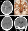
Brain computed tomography (CT) on day 0 showing diffuse subarachnoid hemorrhage (Fisher grade 3) (A). CT angiography on day 0 showing an abnormal cluster of blood vessels at the choroidal point of the anterior choroidal artery (AchA) (arrow, B) and the P2 segment (arrowhead), respectively. A fresh hematoma is visible at the right ambient cistern on axial (C) and coronal (D) CT imaging 1 month after the initial hemorrhage.
FIG. 2
FIG. 2
A: Digital subtraction angiography (DSA) from the right internal carotid artery (ICA) showing the network-like cluster of blood vessels, suggesting a rete mirabile (asterisk) at the choroidal point of the AchA. A small aneurysm (arrow) was detected near the AchA rete mirabile. B: Three-dimensional DSA showing that the AchA rete mirabile (asterisk) leads to the persistence of the temporo-occipital branch. A small aneurysm (arrow) is located at the junction between the rete mirabile and persistent temporo-occipital branch. 1 = posterior communicating artery, 2 = AchA, 3 = uncal branch, 4 = anterior temporal branch, 5 = posterior temporal branch, 6 = occipital branch.
FIG. 3
FIG. 3
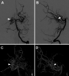
DSA from the vertebral artery, anteroposterior (A) and lateral (B) views, showing the network-like cluster of blood vessels suggesting a rete mirabile (arrowheads) of the posterior cerebral artery (PCA) at the P2 segment. Three-dimensional DSA, anteroposterior (C) and lateral (D) views, showing a rete mirabile (arrowheads) of the PCA at the P2 segment and the peripheral branch of the PCA almost replaced by the AchA.
FIG. 4
FIG. 4
Intraoperative photographs before (A) and after (B) clipping. The asterisk indicates the flow-related aneurysm. Postoperative DSA (C) and three-dimensional DSA (D) showing the disappearance of the flow-related aneurysm (arrows).

Similar articles

References

    1. Kier EL, Conlogue GJ, Zhuang Z. High-resolution computed tomography imaging of the cranial arterial system and rete mirabile of the cat (Felis catus) Anat Rec (Hoboken) 2019;302(11):1958–1967. - PMC - PubMed
    1. Matsoukas S, Fifi JT, Shigematsu T. Lethal subarachnoid haemorrhage in a patient with rete mirabile network of the posterior circulation with associated aneurysm. BMJ Case Rep. 2022;15(2):e245010. - PMC - PubMed
    1. Paschoal EH, Yamaki VN, Júnior FM, Piske RL, Teixeira MJ, Bor-Seng-Shu E. Carotid rete mirabile associated with subarachnoid hemorrhage from intracranial aneurysm: a case report and systematic review. Interv Neuroradiol. 2015;21(1):55–60. - PMC - PubMed
    1. Verma A, Pendharkar H, Prasad C. Rete mirabile of the ICA: report of three cases. Interv Neuroradiol. 2018;24(5):586–590. - PMC - PubMed
    1. Zheng L, Metcalfe R, Bhattacharya JJ. A rete mirabile of the posterior cerebral artery. Embryonic and phylogenetic remnant of choroidal artery origin. Interv Neuroradiol. 2008;14(2):195–202. - PMC - PubMed

LinkOut - more resources