. 2024 Jan 2;43(1):3.
doi: 10.1186/s13046-023-02936-2.

DCAF13 inhibits the p53 signaling pathway by promoting p53 ubiquitination modification in lung adenocarcinoma

Affiliations

DCAF13 inhibits the p53 signaling pathway by promoting p53 ubiquitination modification in lung adenocarcinoma

Shan Wei et al. J Exp Clin Cancer Res. .

Abstract

Background: Lung cancer is a malignant tumor with the highest mortality worldwide. Abnormalities in the ubiquitin proteasome system are considered to be contributed to lung cancer progression with deleterious effects. DDB1 and CUL4 associated factor 13 (DCAF13) is a substrate receptor of the E3 ubiquitin ligase CRL4, but its role in lung cancer remains unknown. In this study, we aimed to investigate the regulatory mechanisms of DCAF13 in lung adenocarcinoma (LUAD).

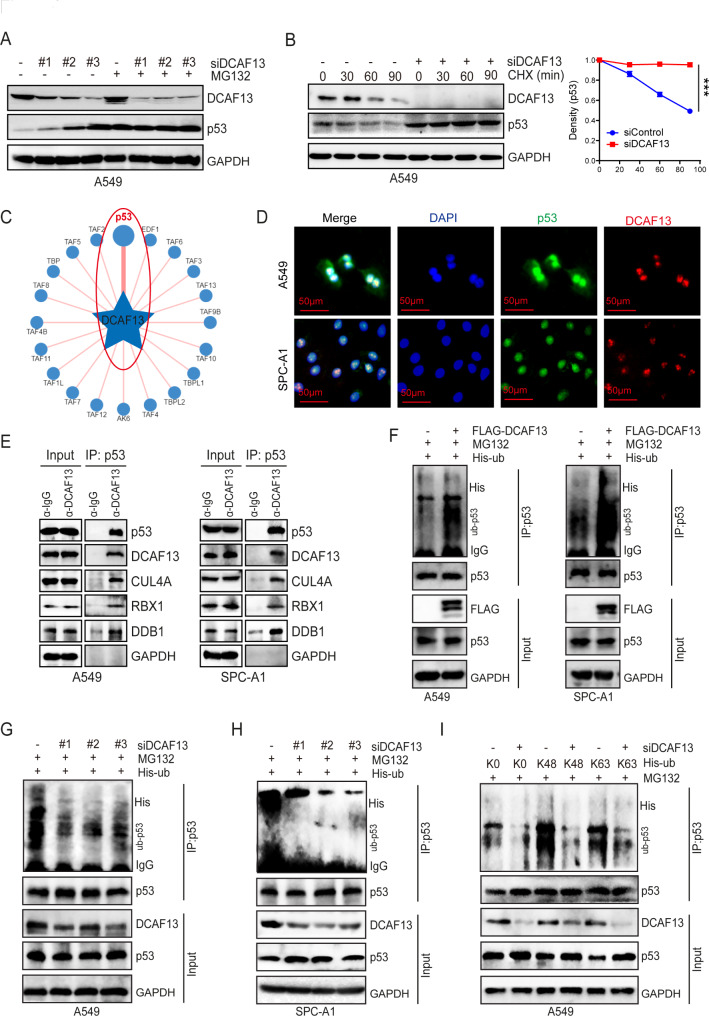
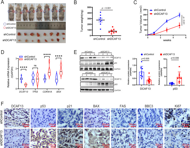
Methods: So as to investigate the effect of DCAF13 on lung adenocarcinoma cell function using in vivo and in vitro. Mechanistically, we have identified the downstream targets of DCAF13 by using RNA-sequencing, as well as ubiquitination assays, co-immunoprecipitation, immunofluorescence, immunohistochemistry and chromatin immunoprecipitation - qPCR experiments.

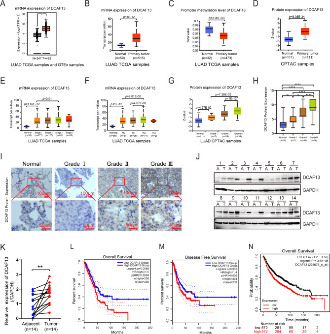
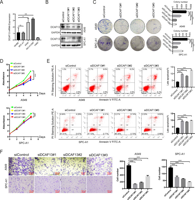
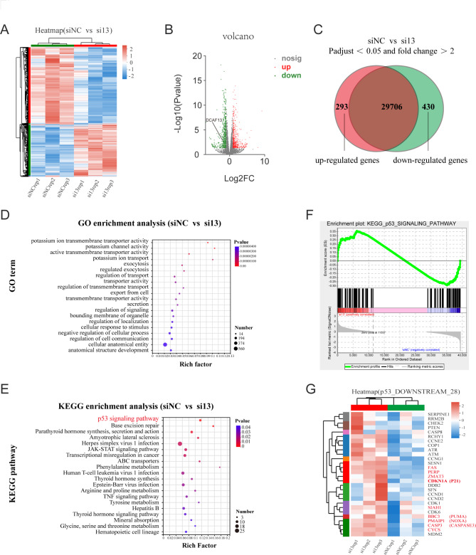
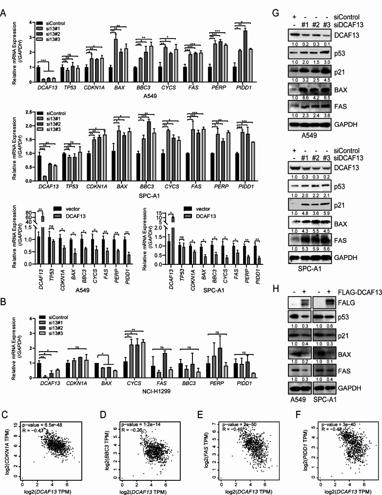
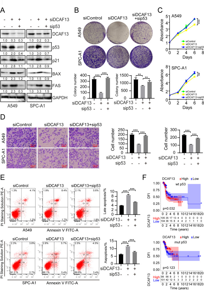
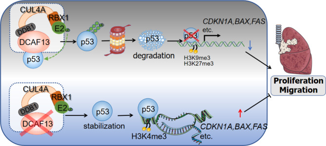
Results: Our findings reveal that DCAF13 is a carcinogenic factor in LUAD, as it is highly expressed and negatively correlated with clinical outcomes in LUAD patients. Through RNA-sequencing, it has been shown that DCAF13 negatively regulates the p53 signaling pathway and inhibits p53 downstream targets including p21, BAX, FAS, and PIDD1. We also demonstrate that DCAF13 can bind to p53 protein, leading to K48-linked ubiquitination and degradation of p53. Functionally, we have shown that DCAF13 knockdown inhibits cell proliferation and migration. Our results highlight the significant role of DCAF13 in promoting LUAD progression by inhibiting p53 protein stabilization and the p53 signaling pathway. Furthermore, our findings suggest that high DCAF13 expression is a poor prognostic indicator in LUAD, and DCAF13 may be a potential therapeutic target for treating with this aggressive cancer.

Conclusions: The DCAF13 as a novel negative regulator of p53 to promote LUAD progression via facilitating p53 ubiquitination and degradation, suggesting that DCAF13 might be a novel biomarker and therapeutical target for LUAD.

Keywords: DDB1 and CUL4 associated factor 13; Lung adenocarcinoma; Protein degradation; Ubiquitination; p53 signaling pathway.


Conflict of interest statement

The authors declare that they have no competing interests.

Figures

Fig. 1
Fig. 1
DCAF13 is highly expressed and associated with poor prognosis in LUAD. A. DCAF13 mRNA expression by GEPIA2. B-G. DCAF13 mRNA expression, promoter methylation level, and protein level by UALCAN. Differences in significance were marked (Mann-Whitney U test). H and I. DCAF13 protein expression levels were detected in a tissue microarray by IHC (Mann-Whitney U test). The scale bar is marked as shown. H, quantification analysis of DCAF13 expression in grade. I, representative DCAF13 immunohistochemical staining. J and K. DCAF13 is highly expressed in fresh clinical LUAD tissue specimens by Western blotting (K, paired t-test). A, Adjacent paraneoplastic tissue; T, Tumor tissue. L-M. Association between DCAF13 mRNA expression and prognosis in TCGA LUAD samples by Kaplan-Meier analysis (log-rank test). L (OS), M (DFS). N. Association between DCAF13 mRNA expression and prognosis in lung cancer by KM-plotter database (log-rank test). Data are presented as mean ± SD. * p < 0.05, ** p < 0.01, *** p < 0.001, and **** p < 0.0001
Fig. 2
Fig. 2
DCAF13 knockdown inhibits LUAD cell growth, migration and apoptosis inhibition in vitro. (A) DCAF13 mRNA expression was detected in LUAD cell lines. The 16HBE cells are normal lung bronchial epithelial cells. A549, SPC-A1, NCI-H1299 cells are LUAD cell lines. H460 cells are human large-cell lung cancer cells. (B) Effective interference of DCAF13 expression by three small interfering RNAs, siDCAF13#1, #2, #3, in A549 and SPC-A1 cells. (C) Cell clone formation experiments were performed in A549 and SPC-A1 cells with DCAF13 knockdown. (D) CCK-8 experiments were performed in A549 and SPC-A1 cells. (E) Flow cytometry was used to analyze the apoptosis rates in A549 and SPC-A1 cells with DCAF13 knockdown, and the percentage of late apoptosis was compared. (F) Cell migration experiments were performed in A549 and SPC-A1 cells with DCAF13 knockdown. Student’s t-test was used in Fig. 2A, C-F. Data presented as mean ± SD, n = 3, * p < 0.05, ** p < 0.01, and *** p < 0.001. Scale bar = 100 μm
Fig. 3
Fig. 3
DCAF13 negatively regulates the p53 signaling pathway. A-C. Heat map, volcano map and venn diagram of differential gene expression in A549 cells followed by DCAF13 knockdown (p-adjust< 0.05 and fold change > 2). D. Bubble map of top 20 pathways for all differential genes by GO enrichment analysis. E. Bubble map of top 20 pathways for all differential genes by KEGG enrichment analysis. F. Gene Set Enrichment Analysis (GSEA) of the p53 signaling pathway. G. Heat map of p53 downstream 28 genes acting core in the Fig. 3F
Fig. 4
Fig. 4
DCAF13 knockdown promotes p53 protein and its downstream target gene expression. (A) The mRNA expression of DCAF13, TP53, CDKN1A, BAX, BBC3, CYCS, FAS, PERP and PIDD1 were detected by RT-qPCR in A549 or SPC-A1 cells transfected with the indicated siRNAs or overexpression plasmids (si13 means siRNA for DCAF13). (B) The mRNA expression of DCAF13, CDKN1A, BAX, BBC3, CYCS, FAS, PERP, and PIDD1 were detected by RT-qPCR in NCI-H1299 cells transfected with siControl or the indicated siRNAs. GAPDH was used as a control. Student’s t-test was used. Data presented as mean ± SD, n = 3, * p < 0.05, ** p < 0.01, and *** p < 0.001. C-F. Correlation between DCAF13 and CDKN1A, BBC3, FAS, PIDD1 mRNA expression in TCGA LUAD and GTEx samples by GEPIA2 (Spearman correlation coefficient). G-H. Protein expression levels of p53, p21, BAX, FAS were analyzed by Western blotting in A549 or SPC-A1 cells transfected with the indicated siRNAs or overexpression plasmids. GAPDH was used as a control
Fig. 5
Fig. 5
DCAF13 knockdown alters histone modifications of p53 target genes. A-D. The p53-RE positions in BAX, CDKN1A, PIDD1, FAS promoter regions and alterations of H3K4me3, H3K9me3 or H3K27me3 on p53-RE by ChIP-qPCR in shControl or shDCAF13 A549 cells. Data were statistically analyzed using Student’s t-test and values are shown as mean ± SD. * p < 0.05, ** p < 0.01, and *** p < 0.001
Fig. 6
Fig. 6
DCAF13 inhibits p53 protein stability via ubiquitination modification. (A) A549 cells were transfected with siDCAF13#1, #2, #3 for 48 h. Before harvest, cells were treated with MG132 (5 μM) for 6 h, cell lysates were subjected to western blotting with indicated antibodies. (B) A549 cells were transfected with siDCAF13#1 for 48 h., Before harvest, cells were treated with 10 mg/mL cycloheximide (CHX) for the indicated time points, and then the cell lysates were used for western blotting. (C) The ubiquitinase and substrate website predicts that p53 is the most potentially substrate specifically recognized by DCAF13 using UbiBrowser database. (D) Immunofluorescence assay was performed to observe the cellular localization of DCAF13 and p53 in A549 and SPC-A1 cells. Scale bar = 50 μm. (E) A549 or SPC-A1 whole cell lysates were immunoprecipitated with protein-A/G agarose beads (mock IgG) or anti-p53 antibody and western blotted with indicated antibodies. Levels of endogenous p53, DCAF13, CUL4A, DDB1 and RBX1 were determined by western blot analysis of cell extracts (input) with indicated antibodies. F-H. A549 or SPC-A1 cells were co-transfected with the indicated siRNA and plasmids for 48 h. Before harvest, cells were treated with MG132 (5 μM) for 6 h. Subsequently, cell lysates were subjected to IP assays with p53 antibody for 12 h and then with protein G beads for 4 h. The mixtures including were ubiquitinated p53 were analyzed by western blot analysis with indicated antibodies. Polyubiquitination level of p53 was detected with the anti-His antibody. I. A549 or SPC-A1 cells were co-transfected with the indicated siRNA and plasmids, including K0 (lysineless), K48 (only K48-linked Ub), and K63 (only K63-linked Ub) as indicated, the following procedure is as shown in Fig. 6F-H
Fig. 7
Fig. 7
DCAF13 knockdown inhibits the LUAD cell growth in vivo. (A) A549 shDCAF13 or shControl cells were implanted into BALB/C nude mice by subcutaneous injection. Photograph was shown as the xenograft tumor with shControl or shDCAF13 A549 cells. Tumors were separated after 5 weeks. (B) Mean tumor weights in mice. (C) Xenograft tumor growth curves in mice. (D) The mRNA expression of DCAF13, TP53, CDKN1A, and BAX in mouse tissues were detected by qPCR. (E) The protein expression of DCAF13 and p53 in mouse tissues were detected by western blot analysis. Data were statistically analyzed using Student’s t-test and values are shown as mean ± SD. **** p < 0.0001. (F) The protein expression of DCAF13, p53, p21, BAX, FAS, BBC3 and Ki67 in mouse tumor tissues were detected by immunohistochemistry assay. Representative images are shown, scale bar = 50 μm
Fig. 8
Fig. 8
Inhibition of p53 could attenuate the effect of DCAF13 in A549 and SPC-A1 cells. (A) Protein expression of p53, p21, BAX, FAS were analyzed by Western blotting in A549 or SPC-A1 cells transfected with the indicated siRNAs. GAPDH was used as a control. (B) Cell clone formation experiments were performed in A549 and SPC-A1 cells transfected with the indicated siRNAs. (C) CCK-8 experiments were performed in A549 and SPC-A1 cells transfected with the indicated siRNAs. (D) Cell migration experiments were performed in A549 and SPC-A1 cells transfected with the indicated siRNAs. (E) Flow cytometry was used to analyze the apoptosis rates in A549 and SPC-A1 cells transfected with the indicated siRNAs. (F) Association between DCAF13 mRNA expression and prognosis in p53 wild-type or mutant type TCGA LUAD samples by Kaplan-Meier analysis. Student’s t-test was used in Fig. 8B-E. Data presented as mean ± SD, n = 3, * p < 0.05, ** p < 0.01, and *** p < 0.001. Scale bar = 100 μm
Fig. 9
Fig. 9
A model diagram of DCAF13 promotes p53 protein ubiquitination degradation via CRL4 complex, which in turn inhibits the p53 signaling pathway, leading to the altered histone modifications of CDKN1A, BAX and FAS, and ultimately promotes LUAD progression

Similar articles

Cited by

References

    1. Sung H, Ferlay J, Siegel RL et al. Global cancer statistics 2020: GLOBOCAN estimates of incidence and mortality worldwide for 36 cancers in 185 countries. CA Cancer J Clin, 2021;71(3):209–249. - PubMed
    1. Siegel RL, Miller KD, Wagle NS, et al. Cancer statistics, 2023. CA Cancer J Clin. 2023;73(1):17–48. doi: 10.3322/caac.21763. - DOI - PubMed
    1. Zappa C, Mousa SA. Non-small cell Lung cancer: current treatment and future advances. Transl Lung Cancer Res. 2016;5(3):288–300. doi: 10.21037/tlcr.2016.06.07. - DOI - PMC - PubMed
    1. Relli V, Trerotola M, Guerra E, et al. Abandoning the notion of Non-small Cell Lung Cancer. Trends Mol Med. 2019;25(7):585–94. doi: 10.1016/j.molmed.2019.04.012. - DOI - PubMed
    1. Miller M, Hanna N. Advances in systemic therapy for non-small cell Lung cancer. BMJ. 2021;375:n2363. doi: 10.1136/bmj.n2363. - DOI - PubMed