

Case Reports
doi: 10.1093/jscr/rjad701.
eCollection 2023 Dec.
Granular cell tumor of the brain: case report and review of literature
Affiliations
- PMID: 38164207
- PMCID: PMC10758243
- DOI: 10.1093/jscr/rjad701
Item in Clipboard
Case Reports
Granular cell tumor of the brain: case report and review of literature
J Surg Case Rep.
.
Abstract
Granular cell tumors are rare tumors that develop from Schwann cells, which are glial cells surrounding neurons of the peripheral nervous system, which serve in the process of myelination. Granular cell tumors are rarely associated with the central nervous system in humans. In this report, we analyze a patient with granular cell tumor and review the current literature.
Keywords: Schwann cells; astrocytoma; granular cell tumors; malignant giant cell.
Published by Oxford University Press and JSCR Publishing Ltd. © The Author(s) 2023.
Conflict of interest statement
None declared.
Figures
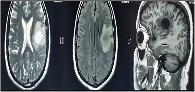
MRI brain with contrast—a heterogeneous irregular rim of an enhancing lesion with a size of 3.6 × 3.4 × 2.4 cm appearing in the left frontal subcortical region, with mild perilesional edema extending into the corona radiata and external capsule.

MRS showed an increased choline, reduced NAA, and prominent lipid/lactate peaks; (Ch/NAA-4.0) (Ch/Cr-1.7).
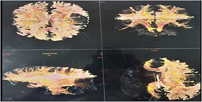
Tractography showing destruction of the left superior longitudinal fasciculus, superior occipito-frontal fasciculus, and partial destruction of the left cortical spinal tract.
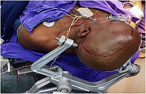
The patient was positioned in the supine position, with the head fixed with a three-pin holder.
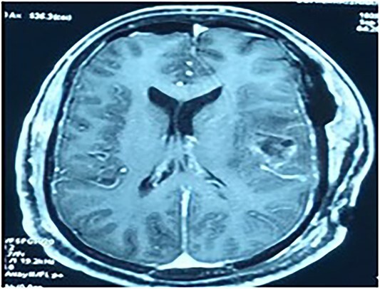
Contrast MRI depicting the tumor cavity and gross total resection.

Section study shows a poorly defined mass composed of sheets of cells separated by thin collagenous bands; cells are round to polygonal with abundant eosinophilic cytoplasm with coarse granules; cell border is distinct at places with a syncytial pattern at some area; nuclei is large and vesicular.
Similar articles
-
Anaplastic astrocytoma with granular cell differentiation: case report and review of the literature.Hum Pathol. 1993 Jul;24(7):805-8. doi: 10.1016/0046-8177(93)90020-h. Hum Pathol. 1993. PMID: 7686529 Review.
-
Clinico-Pathological Features Of Granular Cell Astrocytoma.J Ayub Med Coll Abbottabad. 2021 Oct-Dec;33(Suppl 1)(4):S831-S834. J Ayub Med Coll Abbottabad. 2021. PMID: 35077636
-
Granular cell astrocytoma: report of a case and review of the literature.Clin Neuropathol. 2016 Jul-Aug;35(4):186-93. doi: 10.5414/NP300952. Clin Neuropathol. 2016. PMID: 27125869 Review.
-
Morphologic characterization of spontaneous nervous system tumors in mice and rats.Toxicol Pathol. 2000 Jan-Feb;28(1):178-92. doi: 10.1177/019262330002800123. Toxicol Pathol. 2000. PMID: 10669006 Review.
-
Granular cell tumors in the central nervous system: a report on eight cases and a literature review.Br J Neurosurg. 2016 Dec;30(6):611-618. doi: 10.1080/02688697.2016.1181152. Epub 2016 May 18. Br J Neurosurg. 2016. PMID: 27188824
Cited by
-
Diagnosis and management of oesophageal granular cell tumour: A case report.Exp Ther Med. 2025 Mar 11;29(5):92. doi: 10.3892/etm.2025.12842. eCollection 2025 May. Exp Ther Med. 2025. PMID: 40162053 Free PMC article.
-
Giant Granular Cell Tumor of the Left Thigh, a Rare Case Report and Literature Review.Orthop Res Rev. 2025 Jan 7;17:1-7. doi: 10.2147/ORR.S499488. eCollection 2025. Orthop Res Rev. 2025. PMID: 39801771 Free PMC article.
References
-
- National Cancer Institute . NCI Dictionary of Cancer Terms. n.d.. Retrieved February 19, 2023, from https://www.cancer.gov/publications/dictionaries/cancer-terms/def/granul....
-
- Neelon D, Lannan F, Childs J. Granular Cell Tumor - Statpearls - NCBI Bookshelf. Granular Cell Tumor. n.d.. Retrieved February 19, 2023, from https://www.ncbi.nlm.nih.gov/books/NBK563150/. - PubMed
-
- Gunson DT. Granular Cell Tumour. DermNet. 2018. Retrieved February 19, 2023, from https://dermnetnz.org/topics/granular-cell-tumour.
-
- Vladimir O Osipov, MD . Granular Cell Tumors. Practice Essentials, Etiology and Pathophysiology, Epidemiology. 2022. Retrieved February 19, 2023, from https://emedicine.medscape.com/article/282430-overview.
-
- National Organization for Rare Disorders . Granular Cell Tumor. 2022. Retrieved February 24, 2023, from https://rarediseases.org/gard-rare-disease/granular-cell-tumor/.
Publication types
LinkOut - more resources
Full Text Sources
