

Palliative management of a malignant tracheoesophageal fistula using repeat endobronchial laser debridement and esophageal stenting
- PMID: 38164211
- PMCID: PMC10758224
- DOI: 10.1093/jscr/rjad590
Palliative management of a malignant tracheoesophageal fistula using repeat endobronchial laser debridement and esophageal stenting
Abstract
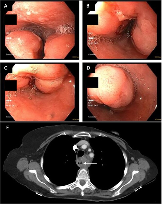
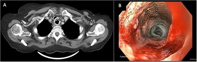
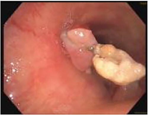
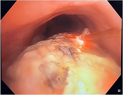
A 71-year-old female presented with progressive dysphagia and unexplained weight loss. Computed tomography and esophagogastroduodenoscopy (EGD) revealed invasive esophageal squamous cell carcinoma, which was initially treated with local radiation and esophageal stenting. Over the next year, the patient experienced multiple symptoms and hospital admissions consistent with a malignant tracheoesophageal fistula, despite negative findings on imaging, bronchoscopy, and EGD. Prophylactic antibiotics were initiated based on symptomatology to prevent septic episodes. Stent erosion into the membranous trachea was eventually observed. Neodymium-yttrium-aluminum-garnet laser bronchoscopy was used periodically to debulk the invading tumor around the stent. A percutaneous endoscopic gastrostomy tube was also inserted to facilitate enteral nutrition and avoid aspiration pneumonia. The patient reported significant improvements in respiratory symptoms following each laser debridement and has progressed well beyond the life expectancy associated with malignant tracheoesophageal fistula.
© Published by Oxford University Press on behalf of JSCR Publishing Ltd. 2023.
Conflict of interest statement
None declared.
Figures
Similar articles
-
Treatment of malignant tracheoesophageal fistula.Thorac Surg Clin. 2014 Feb;24(1):117-127. doi: 10.1016/j.thorsurg.2013.09.006. Epub 2013 Nov 9. Thorac Surg Clin. 2014. PMID: 24295667 Review.
-
Comparative study of esophageal stent and feeding gastrostomy/jejunostomy for tracheoesophageal fistula caused by esophageal squamous cell carcinoma.PLoS One. 2012;7(8):e42766. doi: 10.1371/journal.pone.0042766. Epub 2012 Aug 13. PLoS One. 2012. PMID: 22912737 Free PMC article.
-
Endobronchial management of benign, malignant, and lung transplantation airway stenoses.Ann Thorac Surg. 1995 Jun;59(6):1417-22. doi: 10.1016/0003-4975(95)00216-8. Ann Thorac Surg. 1995. PMID: 7539606
-
Tracheal penetration and tracheoesophageal fistula caused by an esophageal self-expanding metallic stent.Case Rep Pulmonol. 2014;2014:567582. doi: 10.1155/2014/567582. Epub 2014 Sep 3. Case Rep Pulmonol. 2014. PMID: 25276461 Free PMC article.
-
[Interventional treatment of tracheoesophageal/bronchoesophageal fistulas].Chirurg. 2019 Sep;90(9):710-721. doi: 10.1007/s00104-019-0988-z. Chirurg. 2019. PMID: 31240352 Review. German.
References
-
- Balazs A, Kupcsulik PK, Galambos Z. Esophagorespiratory fistulas of tumorous origin. Non-operative management of 264 cases in a 20-year period. Eur J Cardiothorac Surg 2008;34:1103–7. - PubMed
-
- Goh KJ, Lee P, Foo AZX, et al. Characteristics and outcomes of airway involvement in esophageal cancer. Ann Thorac Surg 2021;112:912–20. - PubMed
Publication types
LinkOut - more resources
Full Text Sources
