Choice of Nanoparticles for Theranostics Engineering: Surface Coating to Nanovalves Approach
- PMID: 38164501
- PMCID: PMC10750116
- DOI: 10.7150/ntno.89768
Choice of Nanoparticles for Theranostics Engineering: Surface Coating to Nanovalves Approach
Abstract
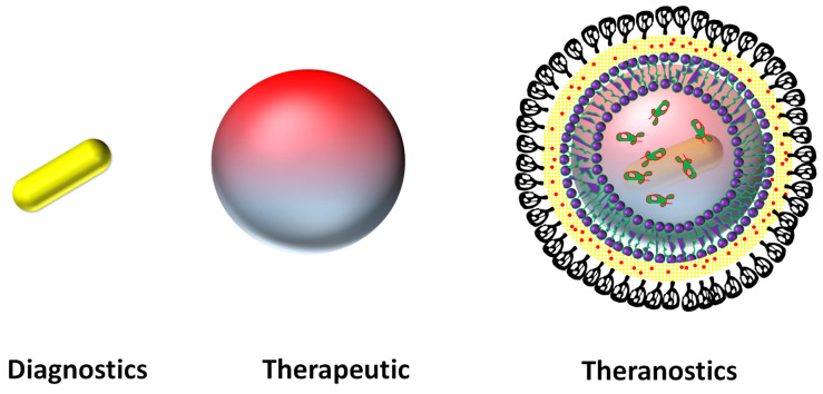
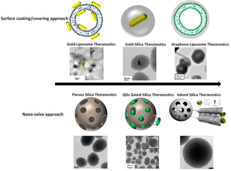
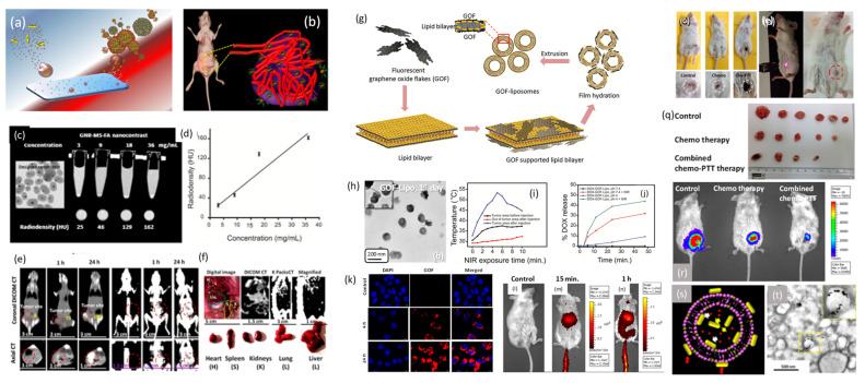
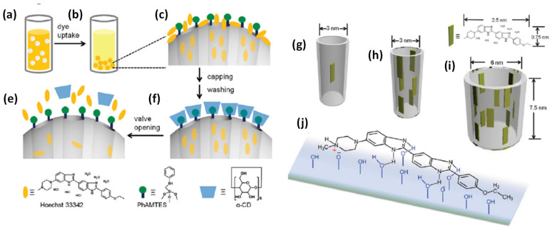
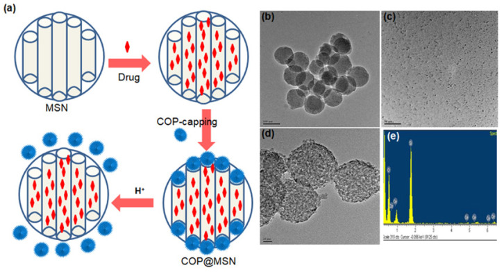
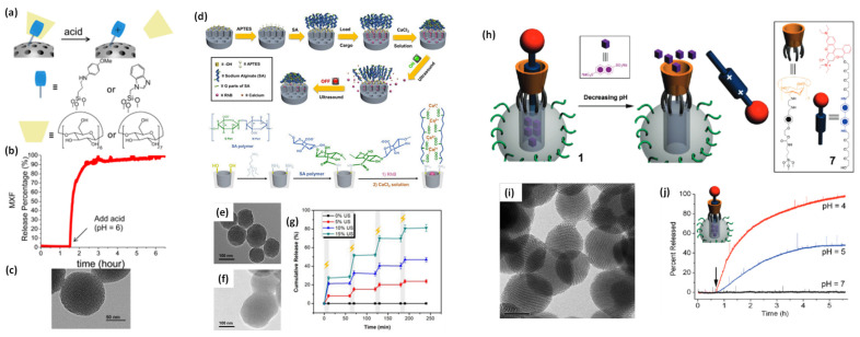
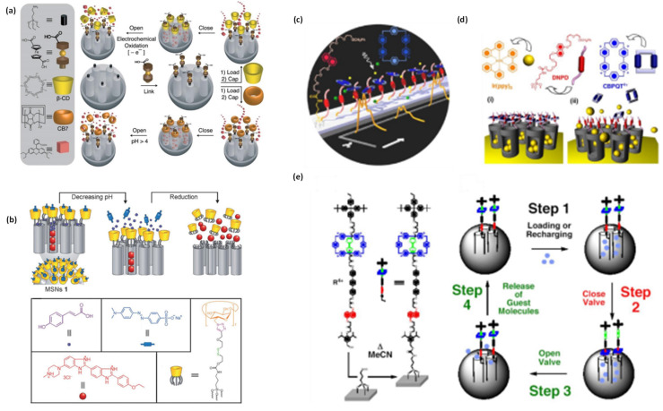
Surface engineered nanoparticles (metallic and nonmetallic) have gained tremendous attention for precise imaging and therapeutics of cell/tumors at molecular and anatomic levels. These tiny agents have shown their specific physicochemical properties for early-stage disease diagnosis and cancer theranostics applications (imaging and therapeutics by a single system). For example, gold nanorods (AuNRs) demonstrate better photothermal response and radiodensity for theranostics applications. However, upon near infrared light exposure these AuNRs lose their optical property which is characteristic of phototherapy of cancer. To overcome this issue, silica coating is a safe choice for nanorods which not only stabilizes them but also provides extra space for cargo loading and makes them multifunctional in cancer theranostics applications. On the other hand, various small molecules have been coated on the surface of nanoparticles (organic, inorganic, and biological) which improve their biocompatibility, blood circulation time, specific biodistribution and tumor binding ability. A few of them have been reached in clinical trials, but, struggling with FDA approval due to engineering and biological barriers. Moreover, nanoparticles also face various challenges of reliability, reproducibility, degradation, tumor entry and exit in translational research. On the other hand, cargo carrier nanoparticles have been facing critical issues of premature leakage of loaded cargo either anticancer drug or imaging probes. Hence, various gate keepers (quantum dots to supramolecules) known nanovalves have been engineered on the pore opening of the cargo systems. Here, a review on the evolution of nanoparticles and their choice for diagnostics and therapeutics applications has been discussed. In this context, basic requirements of multifunctional theranostics design for targeted imaging and therapy have been highlighted and with several challenges. Major hurdles experienced in the surface engineering routes (coating to nanovalves approach) and limitations of the designed theranostics such as poor biocompatibility, low photostability, non-specific targeting, low cargo capacity, poor biodegradation and lower theranostics efficiency are discussed in-depth. The current scenario of theranostics systems and their multifunctional applications have been presented in this article.
© The author(s).
Conflict of interest statement
Competing Interests: The authors have declared that no competing interest exists.
Figures
Similar articles
-
Engineered Liposomes in Interventional Theranostics of Solid Tumors.ACS Biomater Sci Eng. 2023 Aug 14;9(8):4527-4557. doi: 10.1021/acsbiomaterials.3c00510. Epub 2023 Jul 14. ACS Biomater Sci Eng. 2023. PMID: 37450683 Review.
-
Effective Distribution of Gold Nanorods in Ordered Thick Mesoporous Silica: A Choice of Noninvasive Theranostics.ACS Appl Mater Interfaces. 2023 Oct 11;15(40):47615-47627. doi: 10.1021/acsami.3c06108. Epub 2023 Oct 2. ACS Appl Mater Interfaces. 2023. PMID: 37782885
-
Surface Engineering of Nanoparticles toward Cancer Theranostics.Acc Chem Res. 2023 Jul 4;56(13):1766-1779. doi: 10.1021/acs.accounts.3c00122. Epub 2023 Jun 14. Acc Chem Res. 2023. PMID: 37314368
-
Lipid Nanoparticles for Brain Tumor Theranostics: Challenges and Status.Bioconjug Chem. 2024 Sep 18;35(9):1283-1299. doi: 10.1021/acs.bioconjchem.4c00293. Epub 2024 Aug 29. Bioconjug Chem. 2024. PMID: 39207940 Review.
-
Molecular Engineering of Near-Infrared Light-Responsive BODIPY-Based Nanoparticles with Enhanced Photothermal and Photoacoustic Efficiencies for Cancer Theranostics.Theranostics. 2019 Jul 9;9(18):5315-5331. doi: 10.7150/thno.34418. eCollection 2019. Theranostics. 2019. PMID: 31410217 Free PMC article.
Cited by
-
Roadmap on magnetic nanoparticles in nanomedicine.Nanotechnology. 2024 Nov 5;36(4):042003. doi: 10.1088/1361-6528/ad8626. Nanotechnology. 2024. PMID: 39395441 Free PMC article. Review.
-
Exploring the role of strontium-based nanoparticles in modulating bone regeneration and antimicrobial resistance: a public health perspective.RSC Adv. 2025 Apr 7;15(14):10902-10957. doi: 10.1039/d5ra00308c. eCollection 2025 Apr 4. RSC Adv. 2025. PMID: 40196828 Free PMC article. Review.
-
Recent Advances in Photodynamic Therapy: Metal-Based Nanoparticles as Tools to Improve Cancer Therapy.Pharmaceutics. 2024 Jul 12;16(7):932. doi: 10.3390/pharmaceutics16070932. Pharmaceutics. 2024. PMID: 39065629 Free PMC article. Review.
-
Nanoparticles of natural product-derived medicines: Beyond the pandemic.Heliyon. 2025 Feb 19;11(4):e42739. doi: 10.1016/j.heliyon.2025.e42739. eCollection 2025 Feb 28. Heliyon. 2025. PMID: 40083991 Free PMC article. Review.
-
Theranostics: a multifaceted approach utilizing nano-biomaterials.Discov Nano. 2024 Feb 26;19(1):35. doi: 10.1186/s11671-024-03979-w. Discov Nano. 2024. PMID: 38407670 Free PMC article. Review.
References
Publication types
MeSH terms
LinkOut - more resources
Full Text Sources
Medical